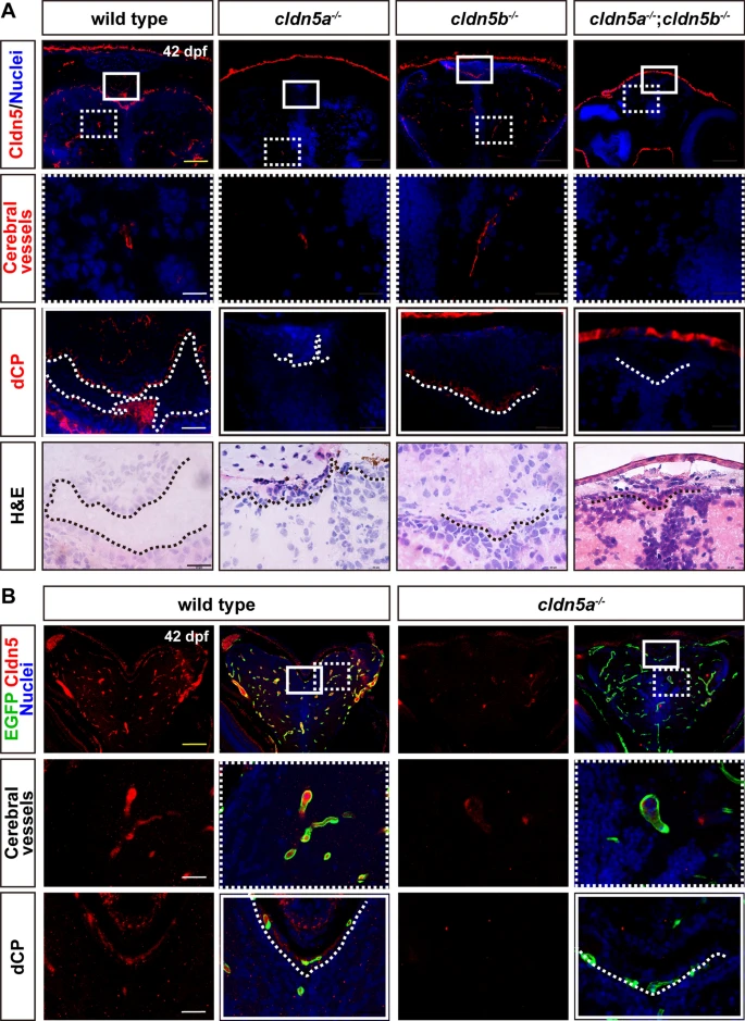
Fig. 1
Expression patterns of Cldn5a and Cldn5b in zebrafish brain. The brain sections of 42 dpf-old zebrafish were stained by an anti-pan Cldn5 antibody. A In wild types, Cldn5s are not only expressed in cerebrovascular ECs but also the dCP epithelium. In cldn5a-/-, Cldn5b expresses only in a few cerebrovascular ECs but not in dCP. In cldn5b-/-, Cldn5a is strongly expressed in both cerebrovascular ECs and dCP. In cldn5a-/-;cldn5b-/-, no specific signals could be detected in the brain. Serial sections were stained with HE to show the histology of dCP. B Tg(kdrl:EGFP) zebrafish was introduced to help to determine the endothelial Cldn5 expression. In cldn5a-/-, Cldn5b is only expressed within a few cerebral vessels but not in dCP epithelium. White rectangles or dashed rectangles indicate the enlarged regions of dCP, or cerebral vessels respectively which are shown in the lower panels with high magnification. White dotted lines or black dotted lines show the continuous CP epithelium. n?=?3 fishes analyzed per group. Scale bars, 100 ?m in yellow or 20 ?m in black or white

