Image
Figure Caption
Figure 1
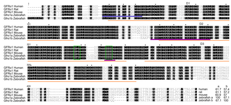
Figure 1. GFRα1 predicted amino acid sequence alignment. Numbers at the top left of the sequences indicate amino acid positions, dashes indicate deletions and black boxes indicate shared sequences. The three cysteine-rich domains (D1–D3) (orange lines), 28 cysteine residues (*) (plus 2 in the terminal region) and two triplets (MLF and RRR) (green boxes) are highly conserved among humans, rodents and zebrafish. At the end of the alignment are the percentage identity values of zebrafish Gfrα1a and Gfrα1b in relation to the other corresponding sequences. The blue line indicates the amino acid sequence recognized by the zebrafish Gfrα1a antibody used in this study; the purple line indicates the putative motifs critical for binding to GDNF.
Acknowledgments
This image is the copyrighted work of the attributed author or publisher, and
ZFIN has permission only to display this image to its users.
Additional permissions should be obtained from the applicable author or publisher of the image.
Full text @ Cells

