
Image
|
Figure Caption
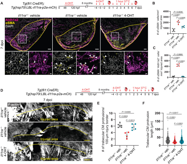
Fig. 7. Il-11 signaling in endothelial cells allows CM repopulation after cardiac injury.