Figure 5
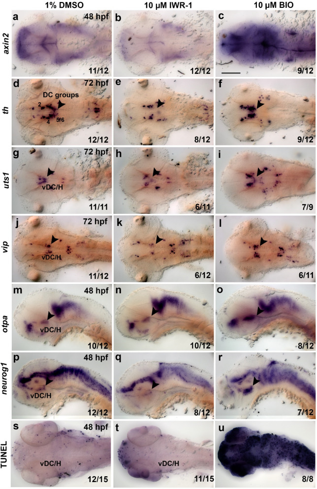
Pharmacological inhibition and activation of Wnt/?-catenin signaling affect neurogenesis of hypothalamic neurons. (a?r) Expression analysis of axin2 (a?c), th (d?f), uts1 (g?i), vip (j?l), otpa (m?o) and neurog1 (p?r) by whole mount in situ hybridization and detection of cell death by the TUNEL assay (s?u) in embryos treated with DMSO (control), IWR-1 or BIO as indicated at top. All embryos were treated from 18 to 42 hpf and fixed at stages indicated in left column of image panels. (a?l) Dorsal and (m?r) lateral views of larval heads, images are Z-projections of image stacks. The arrowheads point to differences in marker gene expression within the ventral diencephalon and hypothalamus. Scale bar in (c) is 100 Ám for all images. Numbers N/N in bottom right corner of each image indicate number of representative phenotypes as shown in image versus total embryos analyzed for this condition. H hypothalamus, vDC ventral diencephalon.

