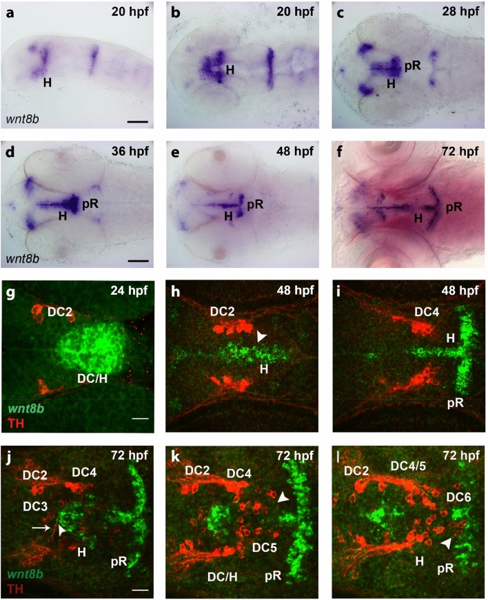
Figure 1
Expression of wnt8b in the ventral diencephalon and hypothalamus and in relation to dopaminergic neurons. (a–f) Expression of wnt8b in embryos as detected by whole mount in situ hybridization at stages as indicated. Lateral (a) and dorsal (b–f) views of heads of embryos, images generated from Z-projections of image stacks. Scale bars in (a) and (d) are 100 µm for (a–f). (g–l) Expression of wnt8b detected by fluorescent in situ hybridization (green) in relation to TH-immunoreactive DA neurons detected by immunofluorescence (red) in embryos at stages as indicated. Dorsal views of the ventral diencephalon/hypothalamus region. Confocal image stacks were recorded and images show single optical sections of Z-stacks containing TH-immunoreactive cells. DA neuron groups DC2 and DC4 of the ventral diencephalon and DC5 and DC6 of the hypothalamus are labeled. (h,i). Two optical sections of a single 48 hpf embryo at 13.97 µm distance from dorsal (h) to ventral (i). (j–l) Three optical section of a single 72 hpf embryo at (j–k) 13.97 µm and (k–l) 5.08 µm distances, with the dorsalmost section shown in (j). Scale bar in (g) is 20 µm for (g–l). (g–l) Z-stacks are included as video files in Supplementary Information as Supplementary Video 1 (g), Supplementary Video 2 (h–i), and Supplementary Video 3 (j–l). Scale bar in (g) is 20 µm for (g–i) and scale bar in (j) is 20 µm for (j–l). DC diencephalon, H hypothalamus, pR posterior recess.

