FIGURE 4
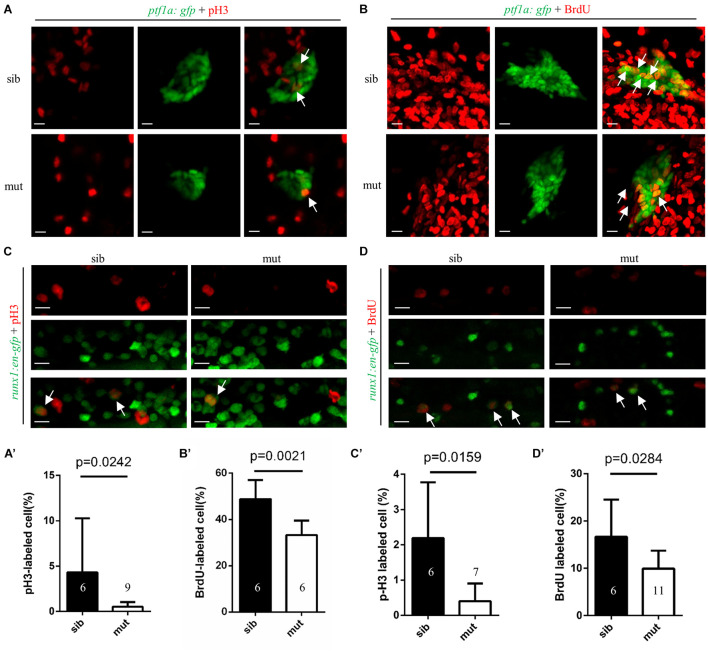
Figure 4. Impaired proliferation of exocrine pancreas progenitors and HSPCs in ltv1?14/?14 mutant. (A,B) Representative confocal images of pH3 immunostaining (A) or BrdU labeling (B) in ltv1?14/?14; ptf1a:gfp mutants and siblings at 2 dpf. (A?,B?) The percentage of pH3+ (A?, siblings, N = 6; mutants, N = 9) or BrdU+ (B?, siblings, N = 6; mutants, N = 6) cells within the ptf1a+ population in ltv1?14/?14 mutants and siblings at 2 dpf. Bars represent means with SD. (C,D) Representative confocal images of pH3 immunostaining (C) and BrdU labeling (D) in ltv1?14/?14; runx1:en-gfp mutants and siblings at 2.5 dpf. (C?,D?) The percentage of pH3+ (C?, siblings, N = 6; mutants, N = 7) and BrdU+ (D?, siblings, N = 6; mutants, N = 11) cells within the runx1+ population in ltv1?14/?14 mutants and siblings at 2.5 dpf. Bars represent means with SD. White arrow: merged cell. Scale bar: 10 ?m.

