IMAGE

Fig. 2.

ID
ZDB-IMAGE-210327-73
Genes
Source
Figures for López-Cuevas et al., 2021
Image
Figure Caption

Fig. 2.

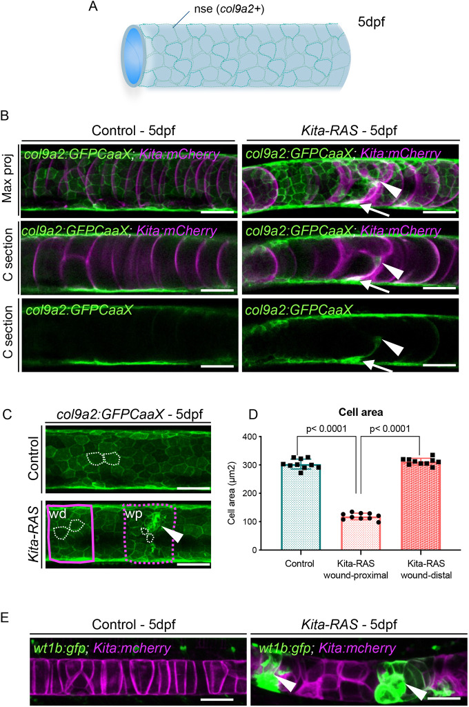
Transformed notochord cells alter the organisation of the sheath layer and activate wound repair mechanisms. (A) Schematic of the notochord and notochord sheath epithelium (nse) at 5 dpf, formed by cells that highly express collagen type IX. (B) Confocal images showing maximum projections (Max proj) and cross sections (C section) of col9a2:GFPCaaX (nse) and kita-mCherry (notochord cells) in control and kita-RAS, at 5 dpf. In kita-RAS, a ‘scar’ region within the notochord (arrowheads) expresses col9a2, and shows connectivity with the nse (arrows). (C) The areas (white dashed lines) of notochord sheath cells were analysed in controls and within two regions of kita-RAS expressing col9a2:GFPCaaX: proximal (wp; magenta dashed line) and distal (wd; magenta solid line) to the wound (arrowhead). (D) Cell area quantification of each group (ten cells were measured for each group and region, and n=10 fish per group). Nested one-way ANOVA and Tukey's multiple comparisons test were used for statistical analysis. Data are mean±s.d. P-values are indicated when significant (P<0.05). (E) Maximum projections from confocal images showing expression of wt1b:gfp in the wounded regions (arrowheads) of kita-RAS. w1b is not expressed in controls. Scale bars: 50 µm.

Figure Data
Acknowledgments
This image is the copyrighted work of the attributed author or publisher, and ZFIN has permission only to display this image to its users. Additional permissions should be obtained from the applicable author or publisher of the image. Full text @ Dis. Model. Mech.