IMAGE

Fig. 1.

ID
ZDB-IMAGE-210327-72
Source
Figures for López-Cuevas et al., 2021
Image
Figure Caption

Fig. 1.

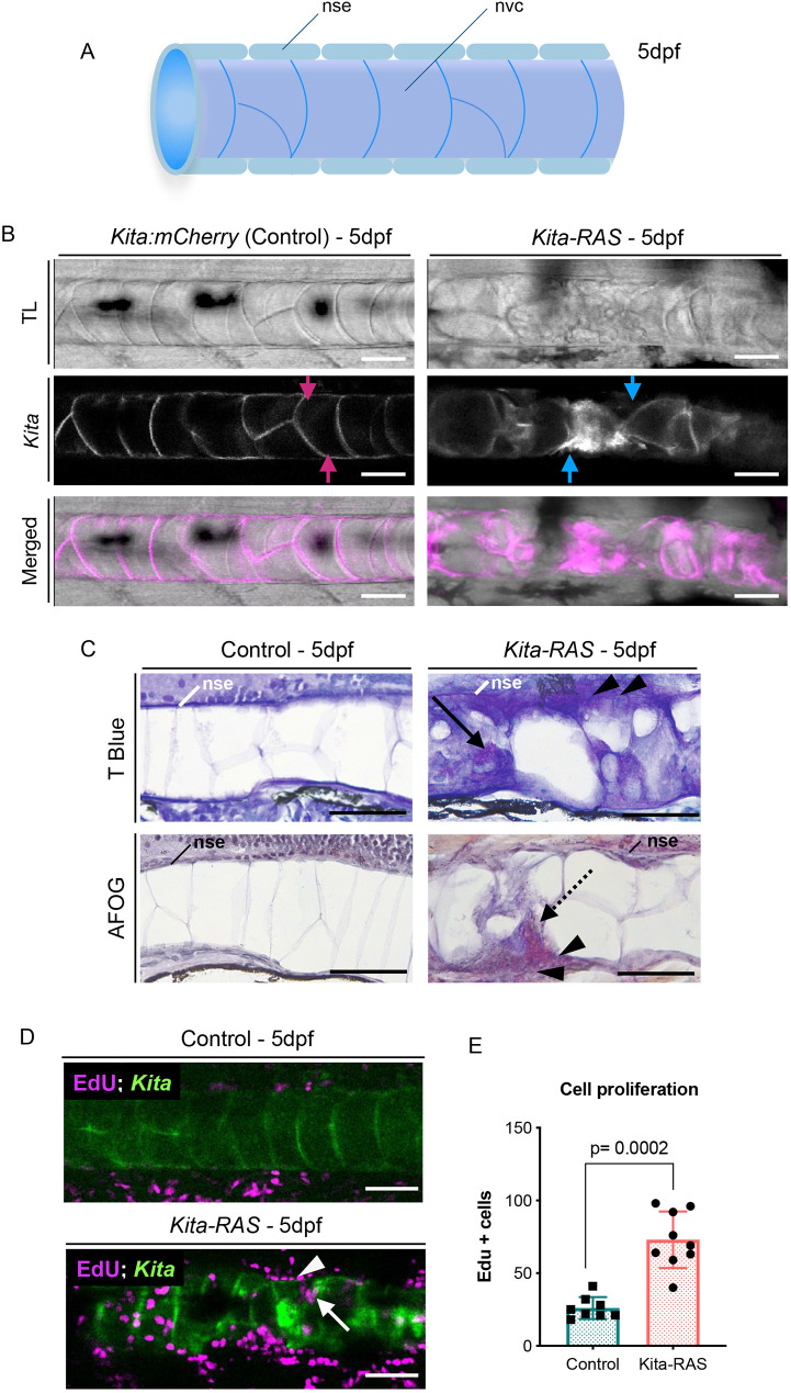
kita drives RAS expression in the notochord,inducing fibrosis and wound-like phenotype. (A) Schematic of the wild-type notochord. The notochord is a rod tube formed by a sealing notochord sheath epithelium (nse) layer that wraps the notochord vacuolated cells (nvc). dpf, days post-fertilisation. (B) Maximum projections from confocal images of control (kita:mCherry) and kita-RAS-mCherry (kita-RAS) at 5 dpf. kita drives expression of the reporter and RAS in the notochord cells (magenta arrows), leading to dramatic changes in the notochord. Gaps between vacuolated notochord cells (blue arrows) are filled with small non-vacuolated cells. (C) Histological sections of 5 dpf control (kita-mCherry) and kita-RAS larvae, stained with Toluidine Blue (T Blue) and AFOG. Control fish show an intact nse. kita-RAS larvae show disruptions of the nse (arrowheads), accumulation of non-vacuolated cells within the notochord (arrow) and fibrous tissue (AFOG, dashed line arrow). (D) Cross section from confocal images of kita (control) and kita-RAS at 5 dpf, treated with EdU from 2 dpf to 4 dpf to show cell proliferation. Note increased proliferation in the notochord sheath (arrowhead) and within wounded areas of the notochord (arrow). (E) Quantification of cell proliferation was performed by counting the number of EdU+ cells in the control (n=8) and kita-RAS (n=9). Nonparametric t-test, post hoc Mann–Whitney test; data are mean±s.d. Scale bars: 50 µm.

Figure Data
Acknowledgments
This image is the copyrighted work of the attributed author or publisher, and ZFIN has permission only to display this image to its users. Additional permissions should be obtained from the applicable author or publisher of the image. Full text @ Dis. Model. Mech.