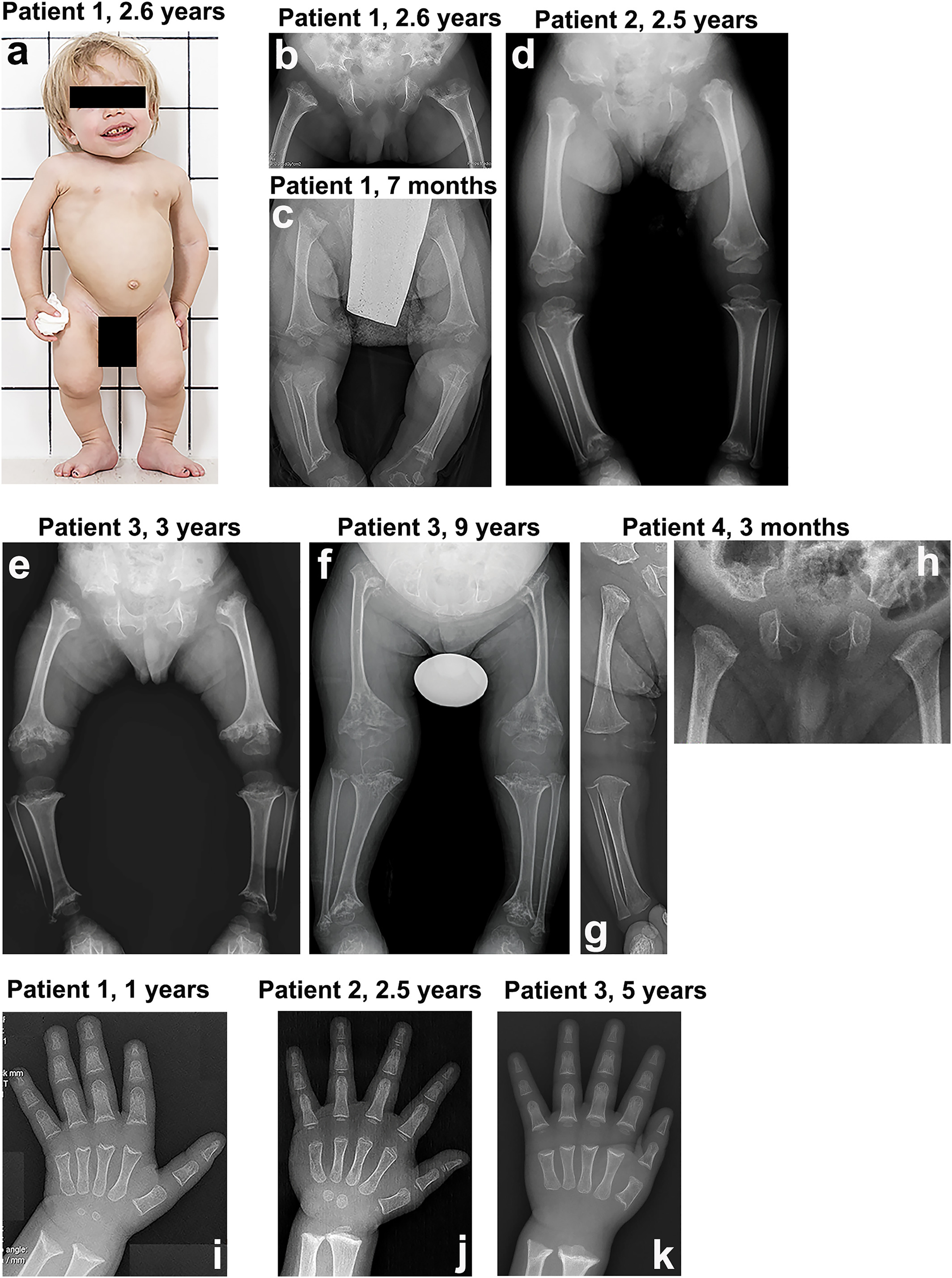
Fig. 1 Clinical and radiological features of the four index patients with spondyloepimetaphyseal dysplasia. (A) Patient 1 at 2.6 years. The clinical features include mild coarseness of facial features with short nose, depressed nasal bridge and hypomineralization of the teeth, severe short stature with short neck, trunk, and limbs, narrow thorax, protruding abdomen, and flexion contractures of the hip and knee joints. Pelvis and lower limbs in patient 1 (B, C), patient 2 (D), patient 3 (E, F), and patient 4 (G, H). The iliac bones are short and broad with serrated iliac crests. The acetabulae are flat and wide with spur‐like projections at the medial and lateral margins. The ischia and pubes are thick with serration at the lateral margin of the ischia. The femora are mildly bowed. The metaphyses of the long bones are wide, cupped, and severely irregular (arrows). The capital femoral epiphyses display significantly delayed ossification, while the other epiphyses of the long bones show normal maturation. The epimetaphyseal changes in patient 3 progressively worsened with age (E, F). Megaepiphyses are found at the knee and ankle. Wrist and hand in patient 1 (I), patient 2 (J), and patient 3 (K). Metaphyseal cupping and irregularity of the radius and ulna are apparent in patients 1 and 3, while those are mild in patient 2. Metaphyseal cupping and irregularity of the metacarpals and proximal phalanges are also found in patients 1 and 3. Delayed carpal bone age is detected in all three patients, but it is more severe in patients 1 and 3. No carpal ossifications are visible in patient 3 at 5 years. Brachydactyly is not apparent in all.
Image
Figure Caption
Acknowledgments
This image is the copyrighted work of the attributed author or publisher, and
ZFIN has permission only to display this image to its users.
Additional permissions should be obtained from the applicable author or publisher of the image.
Full text @ J. Bone Miner. Res.

