IMAGE

Fig 1

ID
ZDB-IMAGE-201121-51
Source
Figures for Schindler et al., 2020
Image
Figure Caption

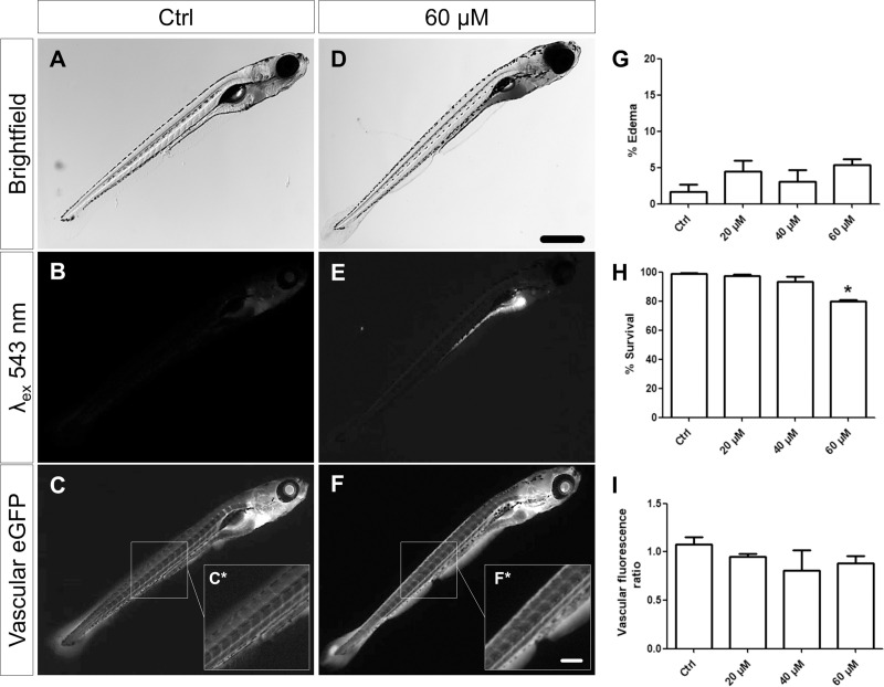
Fig 1 Evaluation of phenotype, autofluorescence, vascular eGFP fluorescence, survival and edema rate of larvae after treatment.

Treated larvae did not develop periocular or pericardial edemas at a significant rate (A, D, G). However, high concentrations of ADR caused an increase in mortality of the larvae (H). All doses of ADR led to a strong accumulation of ADR in the intestinal tract which caused a remarkable autofluorescence over a wide spectrum (B, E). Quantification of the vascular eGFP fluorescence of DBP larvae (Tg(-3.5fabp10a:gc-eGFP) after ADR treatment did not reveal a significant vascular reduction of the eGFP-DBP fusion protein (~68 kDa) (C, F, I). Scale bar in D represents to 200 μm, in F* to 50 μm. *P = 0,03.

Figure Data
Acknowledgments
This image is the copyrighted work of the attributed author or publisher, and ZFIN has permission only to display this image to its users. Additional permissions should be obtained from the applicable author or publisher of the image. Full text @ PLoS One