Fig. 1
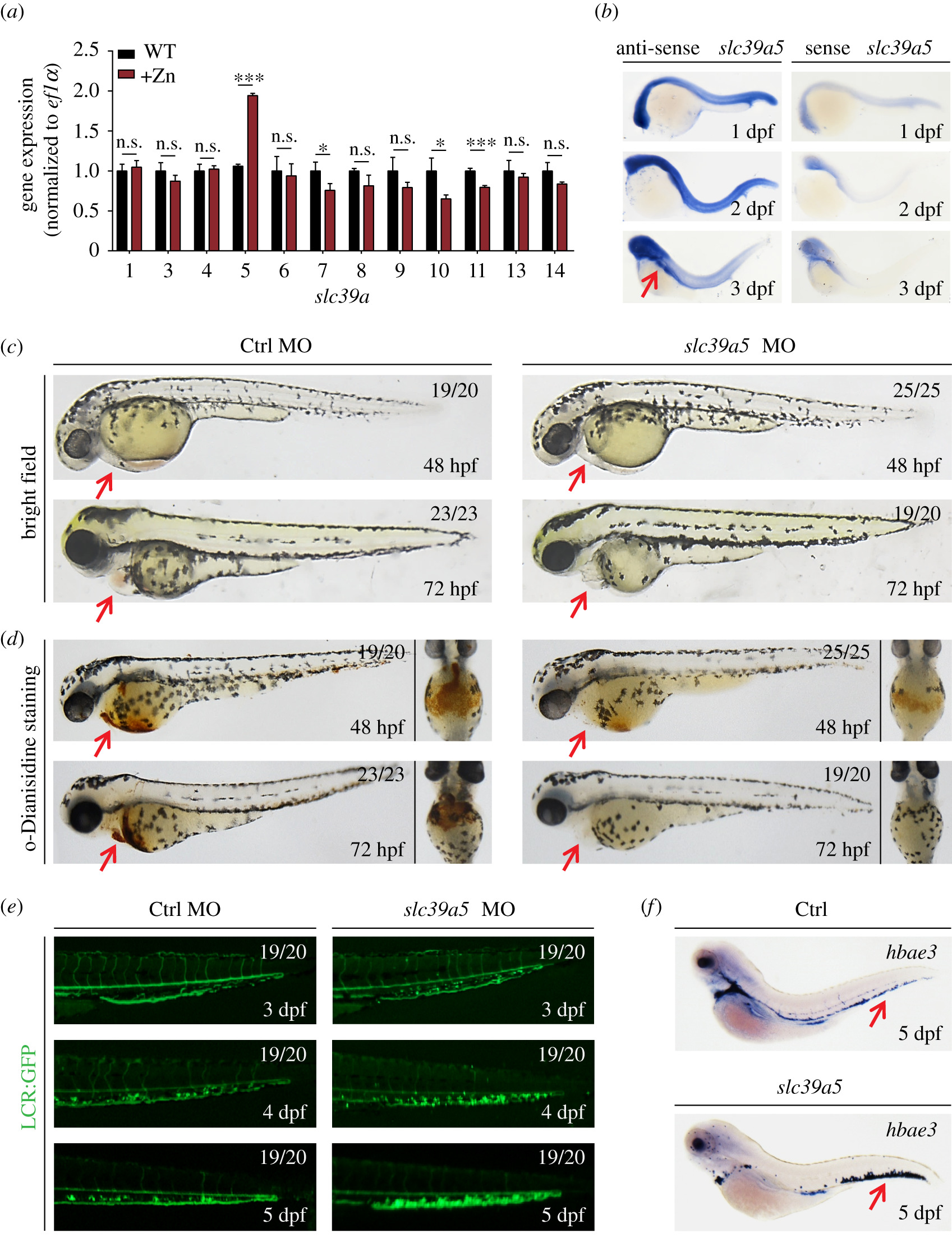
Fig. 1 slc39a5 morphant zebrafish embryos develop cardiac ischaemia and blood cell accumulation in the caudal vein plexus. (a) Quantitative PCR analysis of the 12 indicated slc39a genes measured in wild-type (WT) embryos and embryos treated with 1 mM zinc (+Zn). Note zebrafish lacking slc39a2 and slc39a12. (b) In situ hybridization of WT zebrafish embryos using the antisense and sense (control) slc39a5 probes. Note the concentrated expression in the head and gut region at 3 dpf (red arrow). (c?d) Representative images (c) and o-Dianisidine-stained (d) control and scl39a5 morphant embryos at the indicated developmental stages. The red arrows indicate the heart region. (e) Representative images of GFP fluorescence measured in the caudal region of control and slc39a5 morphant Tg(globinLCR:eGFP) embryos. Note the accumulation of RBCs in the CVP of slc39a5 morphants. (f) Whole-mount in situ hybridization of the RBC marker hbea3 in control and slc39a5 morphants. Note the increased signal in the CVP of the slc39a5 morphant (arrow).

