IMAGE

Figure 2

ID
ZDB-IMAGE-200812-7
Genes
Source
Figures for Ohlebusch et al., 2020
Image
Figure Caption

Figure 2

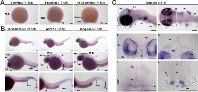
alpl expression during zebrafish embryogenesis. (A) and (B) Spatio-temporal expression pattern of alpl in zebrafish embryos at early segmentation (3–12-somites, A) until long-pec stages (48 hpf, B). (C) Detail images of head regions and of sections stained via ISH for alpl at long-pec stadium. Dashed white lines indicate level of sections shown at higher magnifications (1–4). Scale bar: 100 µm. Ao, dorsal aortic root; Cb, cerebellum; Dc, diencephalon; He, heart tube/heart; Le, lens; Mc, mesencephalon; MHB, midbrain-hindbrain boundary; Ne, nephros; No, notochord; Nt, neural tube; Pf, pectoral fin; Rc, rhombencephalon; Re, retina; Som, somites; Tb, tail bud.

Figure Data
Acknowledgments
This image is the copyrighted work of the attributed author or publisher, and ZFIN has permission only to display this image to its users. Additional permissions should be obtained from the applicable author or publisher of the image. Full text @ Sci. Rep.