IMAGE

Figure 5

ID
ZDB-IMAGE-200812-11
Genes
Source
Figures for Ohlebusch et al., 2020
Image
Figure Caption

Figure 5

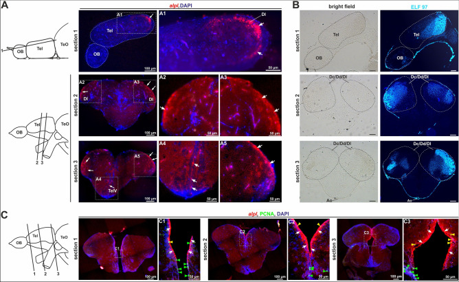
alpl expression pattern (combined with PCNA IF) and Tnap-activity in sections of the adult telencephalon. (A) Sagittal and cross sections of the telencephalic area of an adult zebrafish brain stained with alpl ISH and nuclear staining with DAPI. Signals were detected in the dorsolateral zone of the telencephalon (Dl) and in the ventricular zones, among others the telencephalic ventricle (TelV). The cutting planes of the different sections are indicated through the black lines and marked with numbers. Scale bars: 50 µm or 100 µm (B) ELF 97-staining showed sites of AP-activity within the telencephalon at sites of alpl expression. Scale bars: 100 µm. (C) Cross sections of the telencephalon of an adult zebrafish brain co-stained with alpl ISH (red) and IF for proliferation marker PCNA (green). Blue fluorescence depicts DAPI nuclear staining and gives structural information. C1, C2 and C3 are high resolution images of the respective regions of interest. The alpl ISH signals were indicated with white arrows, positive PCNA IF signals were marked with green arrowheads, and merged signals for both were highlighted using a yellow arrowheads. Scale bars: 50 µm or 100 µm. Ao, dorsal aortic root; Dc/Dd/Dl, central/dorsal/lateral zone of dorsal telencephalon; OB, olfactory bulb; Tel, telencephalon; TeO, Tectum opticum; TeIV, telencephalic ventricles.

Figure Data
Acknowledgments
This image is the copyrighted work of the attributed author or publisher, and ZFIN has permission only to display this image to its users. Additional permissions should be obtained from the applicable author or publisher of the image. Full text @ Sci. Rep.