IMAGE

Fig. 4.

ID
ZDB-IMAGE-200703-14
Source
Figures for Ibrahim et al., 2020
Image
Figure Caption

Fig. 4.

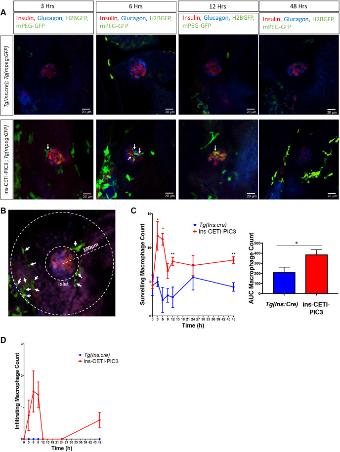
Cytokine-induced embryos have increased macrophage surveillance and infiltration compared to clutch-mate controls. (A) ins-CETI-PIC3 embryos were crossed to the Tg(mpeg:GFP) line that marks macrophages with a GFP tag. CETI-PIC3;ins:cre;Tg(mpeg:GFP) triple-positive embryos were compared to Tg(ins:cre);Tg(mpeg:GFP) control embryos. All embryos were treated with 5 µg/ml doxycycline for different time periods (0-48 h). Infiltrating macrophages are marked with arrows. (B) Schematic for how macrophages were counted. All macrophages within a 100-μm radius of the center of the islet were counted. All macrophages within that radius but outside the islet itself were considered surveilling macrophages (arrows) and all macrophages within the islet were considered infiltrating macrophages. (C) ins-CETI-PIC3 embryos display increased surrounding surveilling macrophages in a time-dependent manner. n=4-5 embryos. *P<0.05, **P<0.01 (parametric t-test). (D) Areas under the curves were calculated to quantify macrophage numbers for multiple embryos and showed that increased infiltrating macrophages were observed in ins-CETI-PIC3 embryos in a time-dependent manner. n=4-5 embryos.

Acknowledgments
This image is the copyrighted work of the attributed author or publisher, and ZFIN has permission only to display this image to its users. Additional permissions should be obtained from the applicable author or publisher of the image. Full text @ Dis. Model. Mech.