Figure 1
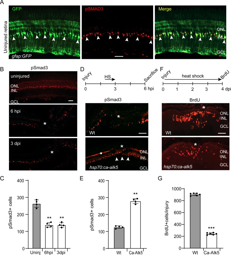
pSmad3 expression in the uninjured and injured retina.
(A) Retinal section from uninjured gfap:GFP fish retina with GFP (green) and pSmad3 (red) immunofluorescence. Arrowheads point to pSmad3 expressing MG. (B) pSmad3 immunofluorescence in uninjured and needle poke injured retina. Asterisk marks injury site. (C) Quantification of data shown in (B). (D) Top diagram shows time line for heat shock treatment after injury and when fish were sacrificed. Bottom panels show pSmad3 immunofluorescence in injured and heat shock-treated Wt and hsp70:ca-Alk5 transgenic fish. Asterisk marks the injury site and arrowheads point to recovery of pSmad3 expression at the injury site in heat shock-treated hsp70:ca-alk5 transgenic fish. White dot in lower panel marks non-specific autofluorescence in the photoreceptor layer. (E) Quantification of data shown in (D). (F) Top diagram is time line for experiment illustrating injury, heat shock treatment and BrdU labelling prior to sacrifice. Lower panels show BrdU immunofluorescence in Wt and hsp70:ca-alk5 fish. Asterisk marks the injury site. (G) Quantification of data presented in (F). Error bars are SD. **p<0.01, ***p<0.001. Scale bar is 50 microns.

