
|
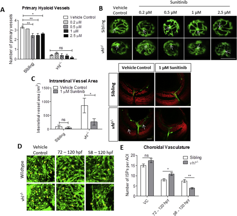
Fig. 5 Sunitinib malate improves hyaloid vessel patterning, reduces retinal neovascularisation and inhibits excessive development of the choriocapillaris in vhl?/? larvae. A) Treatment from 58 hpf does not significantly increase vhl?/? primary hyaloid vessel number at 5 dpf. B) Representative images highlighting an overall improvement of vessel patterning following treatment with sunitinib from 58 hpf. White arrows denote central point from which primary blood vessels are counted. n?=?3, 12 technical replicates per group per experiment. C) Reduction of ectopic intraretinal vasculature following treatment with 1??M sunitinib. White arrows highlighting ectopic intraretinal vessels. Quantification of n?=?5 larvae per treatment group. Scale bar?=?20??m. D) Z-stack projections of the sub-retinal choriocapillaris vasculature (endothelial cells shown in green) of Tg(fli1:EGFP) or vhl?/?(fli1:EGFP) at 120 hpf following treatment for 72 or 48?h as indicated. Yellow arrowheads point to interstitial pillars (ISPs). E) Quantification of the number of interstitial pillars per area of interest (AOI). n?=?10 larvae.
Reprinted from Developmental Biology, 457(2), Ward, R., Ali, Z., Slater, K., Reynolds, A.L., Jensen, L.D., Kennedy, B.N., Pharmacological restoration of visual function in a zebrafish model of von-Hippel Lindau disease, 226-234, Copyright (2019) with permission from Elsevier. Full text @ Dev. Biol.