IMAGE

Fig. 4.

ID
ZDB-IMAGE-200314-78
Genes
Source
Figures for Hau et al., 2020
Image
Figure Caption

Fig. 4.

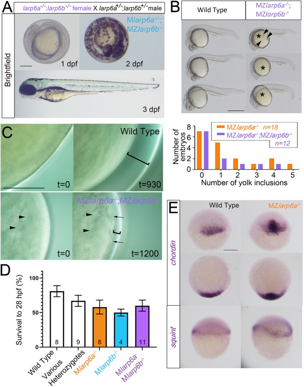
Maternal-zygotic mutant embryos have defective chorion elevation but normal patterning. (A) Three successive stages of a single embryo from a larp6a−/−;larp6b−/− female crossed to a larp6a+/−;larp6b+/− male show that embryonic and chorionic defects do not prevent normal hatching and development. The chorion is tight. All zygotic genotypes appeared similar. Scale bar: 200 μm. (B) Yolk inclusions (arrowheads) in 24 hpf larvae of the indicated genotype, quantified in the graph. The yolk (asterisks) is yellow in double mutants. Scale bars: 500 µm. (C) Timelapse imaging of chorion elevation reveals failure in eggs derived from double mutant females. There is reduced chorion elevation (brackets) and uneven cytoplasm in unactivated egg (arrowheads), and uneven egg surface after activation (arrows). Scale bar: 200 μm. (D) Mild reduction in survival of fertilised eggs between 1 and 28 hpf in lays from mutant females. Subsequent survival did not differ between genotypes and no effect of paternal genotype was observed. Data are mean±s.e.m., number of lays is indicated on each column. (E) In situ hybridisation for the dorsal marker gene chordin and germring marker gene squint show that Mlarp6a−/− embryos are not ventralised compared with wild-type controls at 30% epiboly. Upper panels are lateral views; lower panels are animal views; arrowhead indicates dorsal. Scale bar: 200 µm.

Figure Data
Acknowledgments
This image is the copyrighted work of the attributed author or publisher, and ZFIN has permission only to display this image to its users. Additional permissions should be obtained from the applicable author or publisher of the image. Full text @ Development