IMAGE

Fig. 2

ID
ZDB-IMAGE-200227-4
Source
Figures for Chen et al., 2020
Image
Figure Caption

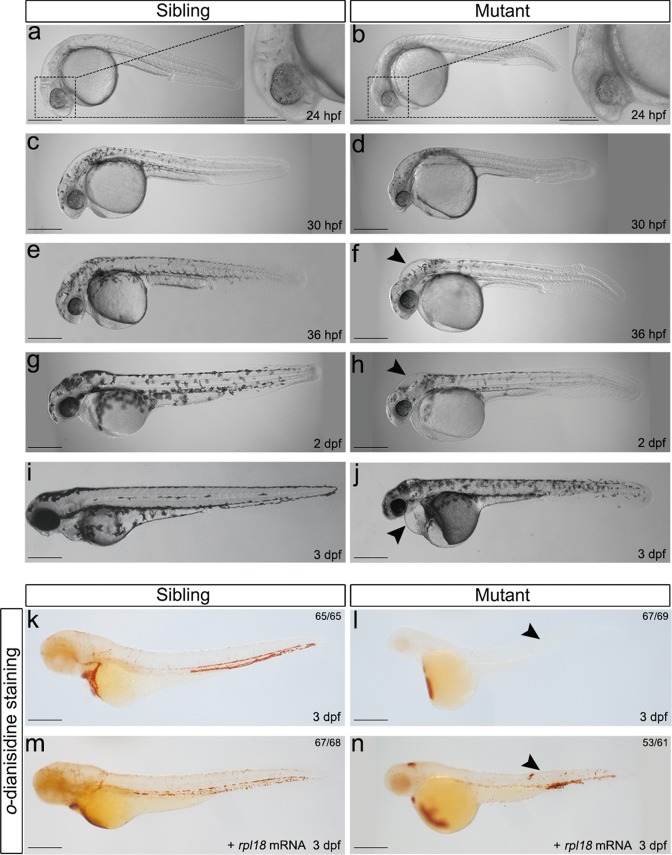
Fig. 2 Rpl18-deficient embryos displayed physical abnormalities and anemia.

aj Phenotypes of wild-type siblings and rpl18−/− mutants (N = 7). At 24 hpf (a, b), aplasia of head region (dashed box) can be seen in mutant. At 30 hpf (c, d), pigmentation was delayed. At 36 hpf (e, f) and 2 dpf (g, h), a smaller head and edema (arrowhead) were present. At 3 dpf (i, j), edema (arrowhead) in heart and shorted tail extension was more apparent. Almost all embryos died at 4 dpf. k, lo-dianisidine staining showed depletion of erythroid cells in rpl18−/ embryos compared with wild-type siblings (arrowheads pointed to the sites of weak o-dianisidine staining, N = 5). m, nrpl18 mRNA (100 pg/embryo) injected to embryos, staining of 3 dpf mutants with o-dianisidine showed a partial recovery (N = 3). All scale bars represent 250 μm.

Figure Data
Acknowledgments
This image is the copyrighted work of the attributed author or publisher, and ZFIN has permission only to display this image to its users. Additional permissions should be obtained from the applicable author or publisher of the image. Full text @ Cell Death Dis.