IMAGE

Fig. 4.

ID
ZDB-IMAGE-191230-1889
Genes
Source
Figures for Liu et al., 2019
Image
Figure Caption

Fig. 4.

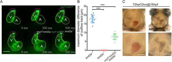
Mechanical behaviours of OFT are deregulated in tnnt2a mutant zebrafish with myocardial tnnt2a supplementation. (A) Representative images of the OFT morphologies at systole and diastole stages of a cardiac cycle. V, ventricle; A, atrium; yellow dotted line, heart outline; white line, OFT diameter; purple dotted line, OFT outline. Scale bar: 100 µm. (B) Statistics of maximum diameter difference of OFT in tnnt2a+/+, tnnt2a−/− and Tg(myl7:tnnt2a); tnnt2a−/−. ****P<0.0001; n=30 per group. Statistical differences were evaluated using Kruskal–Wallis test. (C) Images of the stacked blood cells in the heart chambers of tnnt2a−/− and Tg(myl7:tnnt2a); tnnt2a−/− zebrafish at 72 hpf. Arrows show the stacked red blood cells. Scale bars: 100 µm.

Figure Data
Acknowledgments
This image is the copyrighted work of the attributed author or publisher, and ZFIN has permission only to display this image to its users. Additional permissions should be obtained from the applicable author or publisher of the image. Full text @ Biol. Open