
|
Fig. 6
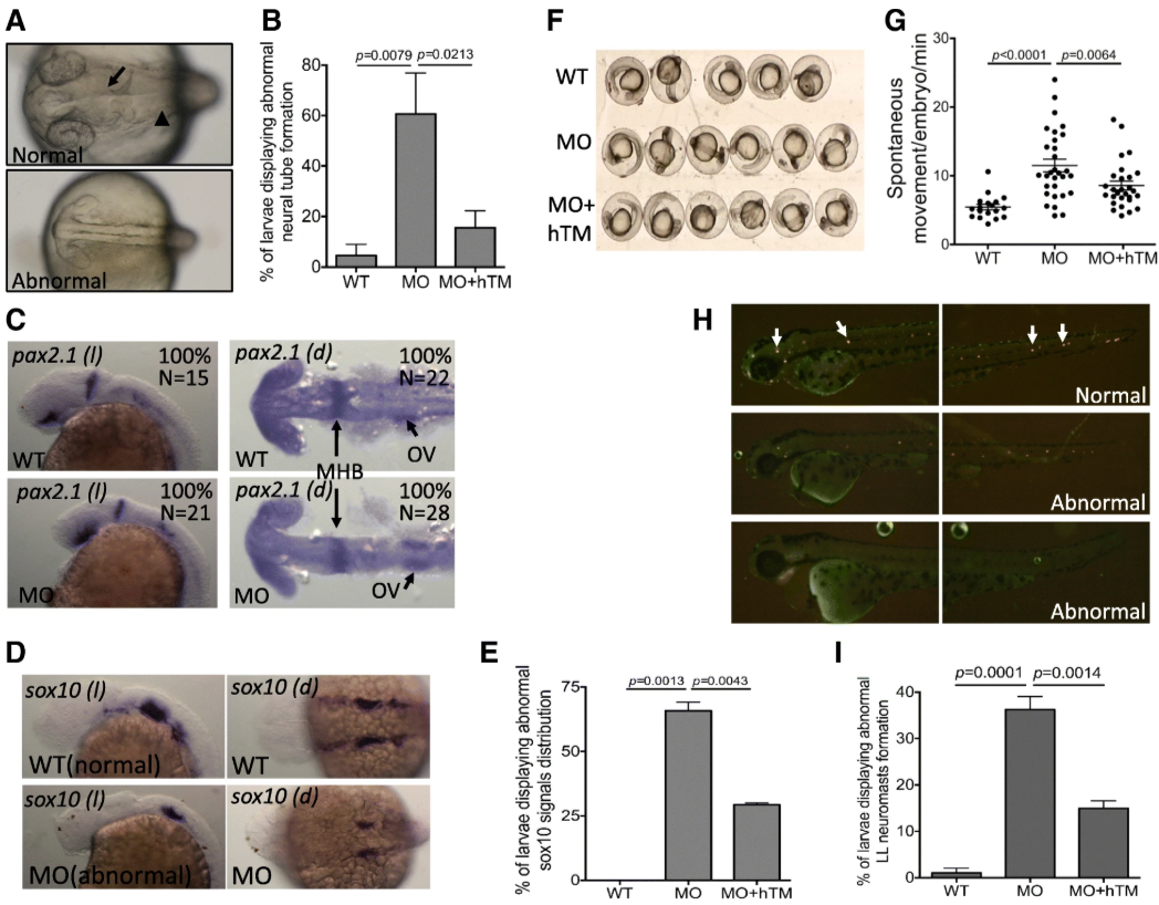
The impact of zTM-b knockdown in neural development. Wild-type embryos, subjected to with/without injecting TM-b MO and rescuing with hTM, were observed and collected at the indicated stages for evaluating the neural tissues development. a, b Embryos at 1 dpf were imaged dorsally under a light dissecting microscope. Those embryos displaying absence or aberrant formation of brain ventricle (arrow) and brain structure (such as mid-hind brain boundary indicated by arrowhead) were categorized as abnormal and quantified. c- e Embryos at 1 dpf were subjected to WISH for the distribution of pax2.1 (a marker of central nervous system) and sox10 (a marker of neural crest cells). Embryos were imaged both laterally ( l) and dorsally ( d) with anterior to the left. e Embryos displaying signal with altered intensity or distribution pattern for sox10, in comparison to wild-type, were categorized as abnormal and quantified. f, g Larval spontaneous movement was examined by video-recording the embryos at 1 dpf for 5 min and counting for the numbers of moving episode for each embryo. Presented here is a representative image of a series of video stills showing the arrangement of examined embryos. h, i The neuromasts of lateral line (arrows) in embryos of 2 dpf were visualized by staining with 4-Di-2-Asp and imaged laterally with anterior to the left. Those embryos displaying absent or aberrantly distributed neuromasts were categorized as abnormal and quantified. Data were collected from at least 3 independent experiments with the total sample numbers of 18–127 for each group in different experiments. WT, wild-type embryos; MO, zTM-b morphants; hTM, human thrombomodulin; OV, Otic vesicles