
|
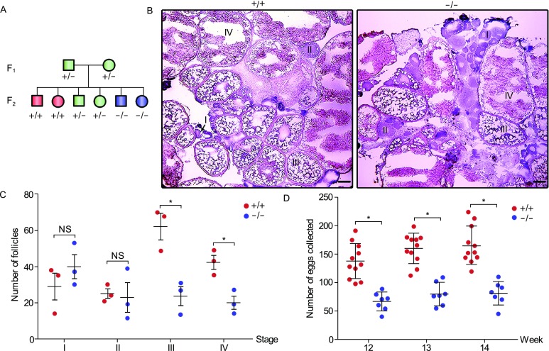
Figure 2
Knockout of sox3 leads to follicle development retardation and a reduced fecundity in female zebrafish. (A) Schematic diagram of heterozygote incrosses. All data are from the progeny derived from heterozygote incrosses. WT, +/+; heterozygotes, +/−; homozygotes, −/−. (B) Histological analysis of adult ovaries of sox3+/+ and sox3−/− by H.E. staining. I, primary growth stage; II, cortical alveolus stage; III, early-vitellogenic stage; IV, late-vitellogenic stage. Scale bar, 50 μm. (C) Statistic analysis of different stages of follicles from 10 sections for each ovary of sox3+/+and sox3−/− (n = 3 fish per genotype). Data represent means ± SEM. T-test was performed. *P < 0.05; **P < 0.01. NS, not significant. (D) Impaired fecundity in sox3−/−. Statistical analysis of the numbers of eggs produced by sox3−/− incrosses or sox3+/+ incrosses at 12-, 13- and 14-week. Data represent means ± SEM. T-test was performed. *P < 0.05