
|
Fig. 5
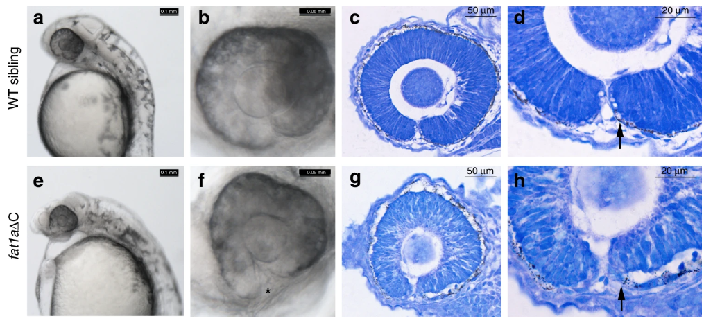
Zebrafish embryos with homozygous alleles of truncated fat1a display coloboma. CRISPR/Cas9-mediated introduction of frame-shift mutations in FAT1 C-terminal resulted in optic fissure closure defects (a and e, scale bar is 0.1 mm). A higher magnification of eye depicting fused margins in WT and unfused margins in homozygous mutant (*, b and f, scale bar is 0.05 mm). Sagittal sections of zebrafish embryos (24–30 hpf) followed by toludene blue staining showing organization of the optic cup (c and g, scale bar is 50 µm). Higher magnification of the optic cup shows morphology of optic fissure margins in WT and homozygous mutant (d, h, scale bar is 20 µm)