Image
Figure Caption
Fig. 1
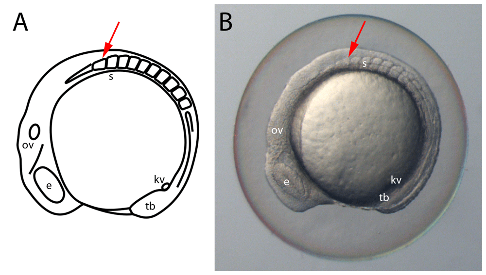
Staging embryos by somite counting.
A schematic (A) and light-microscopy image (B) showing a lateral view of a 10 somite stage zebrafish embryo. The most anterior somite (red arrow) is slightly shorter and broader than more posterior somites. The generation of somites is tightly linked to the overall development of the embryo and therefore allows for accurate aging. e, eye; kv, Kupffer?s vesicle; ov, otic vesicle; s, somites; tb, tail bud.
Acknowledgments
This image is the copyrighted work of the attributed author or publisher, and
ZFIN has permission only to display this image to its users.
Additional permissions should be obtained from the applicable author or publisher of the image.
Full text @ F1000Res

