
|
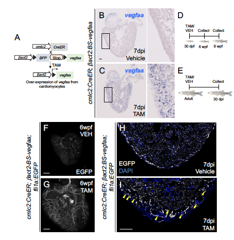
Fig. S2
Transgenic overexpression of vegfaa from cardiomyocytes. (A) Transgenic approach to overexpress vegfaa from cardiomyocytes. (B-C) In situ hybridization for vegfaa on sections from ventricles of cmlc2:CreER; ?act2:BS-vegfaa animals 7 days post incubation (dpi) with vehicle or tamoxifen. Violet staining indicates expression. Boxed region corresponds to the magnified region shown in the adjacent panels. (D-E) Schematic depicting juvenile and adult experiments in Figure 3. (F-G) Tile-scanned images of ventricular surfaces from cmlc2:CreER; ?act2:BS-vegfaa; fli1a:EGFP fish at 6 wpf following incubation with vehicle or tamoxifen at 30 dpf. (H-I) Tile-scanned images of sections from ventricles of cmlc2:CreER; ?act2:BS-vegfaa; fli1a:EGFP animals 7 days after treatment with vehicle or tamoxifen. Yellow arrows indicate nucleated blood cells in the lumen of ectopic blood vessels. (Scale bars 100 ?m)