Fig. 2
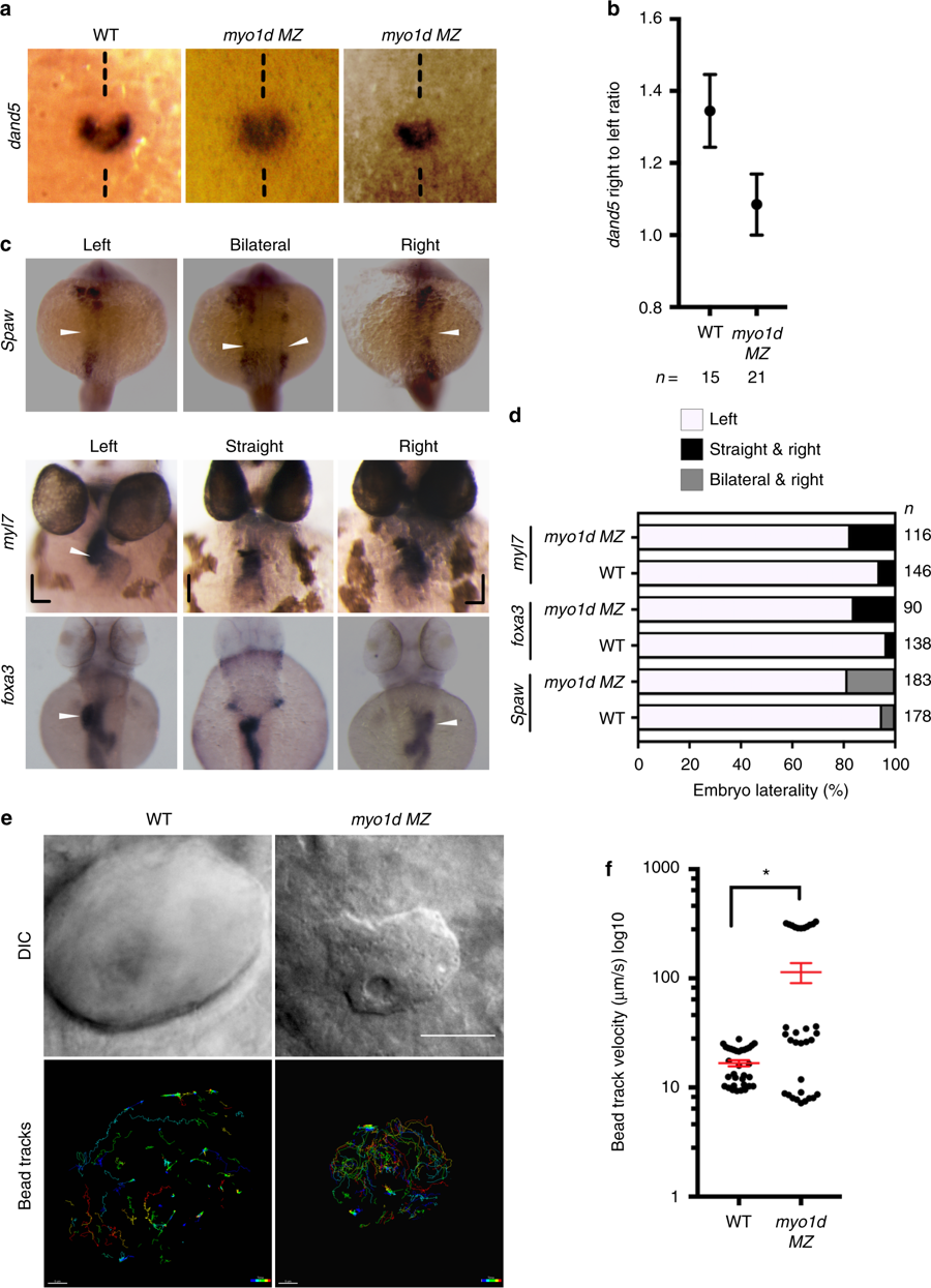
myo1d is necessary for KV function and establishing laterality. a, b Relative abundance of dand5 expression on the right vs left were decreased in myo1d MZ mutants compared to WT embryos at 8S. c, d myo1d MZ mutants displayed laterality defects. Heart and gut looping scored by myl7 and foxa3 expression at 72?hpf respectively, spaw expression at 17S (white arrowhead marks expression). Laterality defects were scored in d. e, f myo1d MZ embryos display altered KV flow as analyzed by tracking 0.2?µ FITC fluorescent microbeads injected into KV lumen. DIC images showing KV morphology of control and myo1d MZ mutants (e). Individual beads were mapped showing unidirectional circular movement were lost in myo1d MZ mutants compared to wild-type embryos. Individual bead movement was shown as temporal color-coded tracks. Graph comparing bead track velocity (mean?±?SEM) in control (n?=?33 tracked beads from three embryos) and myo1d MZ mutant embryos (n?=?33 tracked beads from three embryos) (f). Unpaired t test indicate statistical significance * p?<?0.05. Scale bar, 50?µm

