
|
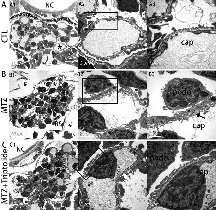
Fig. 3
Triptolide reversed podocyte foot process effacement in MTZ-treated Tg(pod:gal4;UAS:NTR-mCherry) zebrafish. (A) Electron micrograph of normal glomerular vascular loop morphology and foot process development at 6 dpf in wild-type larvae. (B) Glomerular morphology of zebrafish treated with MTZ for 24?h. At low magnifications, an enlarged space outside of Bowman?s capsule was observed, indicating oedema (B1, indicated by #). Extensive foot process effacement was observed at higher magnifications (B3, indicated by arrows). (C) Triptolide prevented most foot process effacement. (n?=?3). *Capillary that was magnified. podo, podocyte; cap, capillary space; BS, Bowman?s space; NC, notochord. Scale bar?=?10 ?m (A1,B1,C1), 2 ?m (A2,B2,C2), 1 ?m (A3,B3,C3).