
Fig. 5
|
Fig. 5
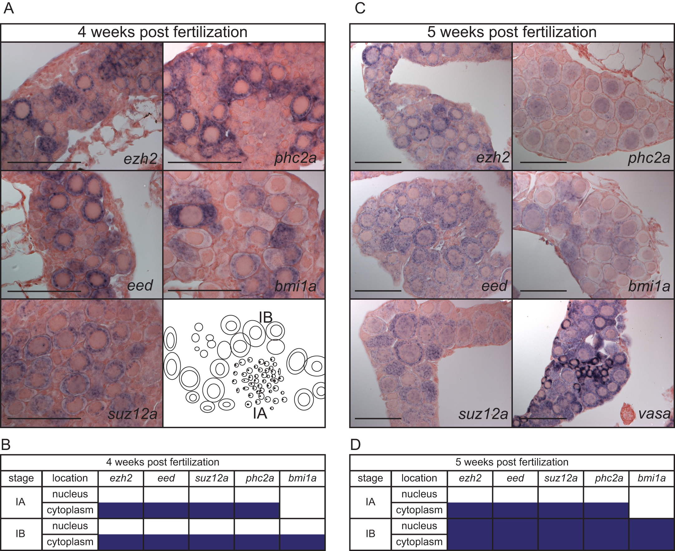
Expression of PcG genes in developing gonads.
(A) Spatio-temporal expression assessed by whole mount in situ hybridization of ezh2, eed, suz12a, phc2a, and bmi1a at 4 weeks post fertilization. (B) Expression patterns of ezh2, eed, suz12a, phc2a, and bmi1a, in gonads at 4 weeks post fertilization. Purple boxes indicate expression of the corresponding gene at that stage of oogenesis. (C) Spatio-temporal expression assessed by whole mount in situ hybridization at 5 weeks post fertilization of ezh2, eed, suz12a, phc2a, bmi1a, and the germ cell marker vasa. (D). Expression of ezh2, eed, suz12a, phc2a, and bmi1a, at 5 weeks post fertilization. Purple boxes indicate expression of the corresponding gene at that stage of oogenesis. Stages of oogenesis are assessed according to Selman et al. [32].