Fig. S6
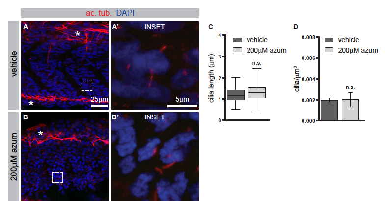
Azumolene does not affect the formation of primary cilia. Related to Figure 6. (A and B) Immunohistochemistry to detect acetylated tubulin (ac. tub.), which marks primary cilia (red), and DAPI-staining to indicate nuclei (blue). Neural axons are also labeled by ac. tub. and are indicated by asterisks. Higher magnification views in A? and B? are indicated by white boxes. (C and D) Quantification of the lengths of the ciliary axoneme or the number of cilia per micron cubed. In C, boxes indicate 50th percentile, whiskers indicate the minimum and maximum values, and median is indicated by the line. In D, data are represented as mean ąSEM. n.s., not significant.
Reprinted from Developmental Cell, 45(4), Klatt Shaw, D., Gunther, D., Jurynec, M.J., Chagovetz, A.A., Ritchie, E., Grunwald, D.J., Intracellular Calcium Mobilization Is Required for Sonic Hedgehog Signaling, 512-525.e5, Copyright (2018) with permission from Elsevier. Full text @ Dev. Cell

