
Fig. 1
|
Fig. 1
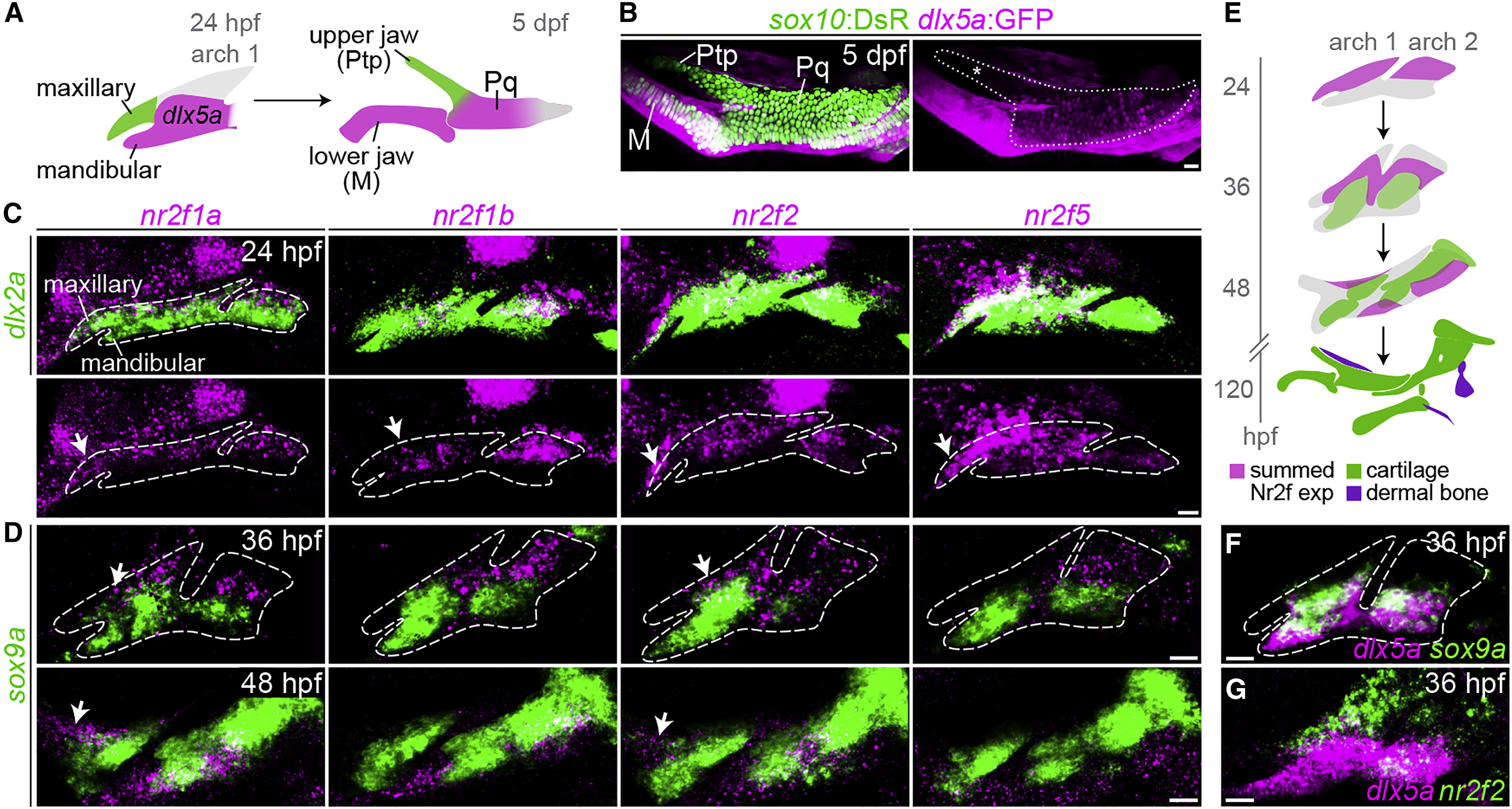
Enrichment of Nr2f Genes in the Maxillary Domain
(A) The pterygoid process (Ptp) of the larval upper jaw derives from cells in the maxillary prominence (green), whereas the body of the palatoquadrate cartilage (Pq) derives from a large dlx5a+ pre-cartilaginous condensation (magenta) that also gives rise to the lower-jaw Meckel's cartilage (M).
(B) dlx5a:GFP staining (magenta) in the body of the Pq but not the Ptp (asterisk) confirms their distinct origins. Chondrocytes are labeled by sox10:DsRed (green). The dotted line demarcates the full Ptp-Pq element.
(C) Double-fluorescence in situ hybridizations for four Nr2f genes (magenta) at 24 hpf, with dlx2a (green) marking arch NCCs. Images are representative maximum-intensity projections. Also see Figure S1 for quantitative expression data and Table S3 for in situ probe information.
(D) Nr2f expression (magenta) is largely excluded from nascent sox9a+ pre-chondrocytes (green) at 36 and 48 hpf. Top images are single z slices; bottom images are maximum-intensity projections.
(E) Summed Nr2f expression domains in the first and second arches and relative to developing cartilage elements.
(F) dlx5a co-localizes with sox9a expression (single z slice).
(G) Co-staining shows a sharp boundary between maxillary nr2f2 and mandibular dlx5a expression (maximum-intensity projection).
White arrows in (C and D) indicate maxillary expression. Dashed lines in (C, D, and F) reflect approximate arch boundaries. Scale bars, 20 ?m.
Reprinted from Developmental Cell, 44(3), Barske, L., Rataud, P., Behizad, K., Del Rio, L., Cox, S.G., Crump, J.G., Essential Role of Nr2f Nuclear Receptors in Patterning the Vertebrate Upper Jaw, 337-347.e5, Copyright (2018) with permission from Elsevier. Full text @ Dev. Cell