
|
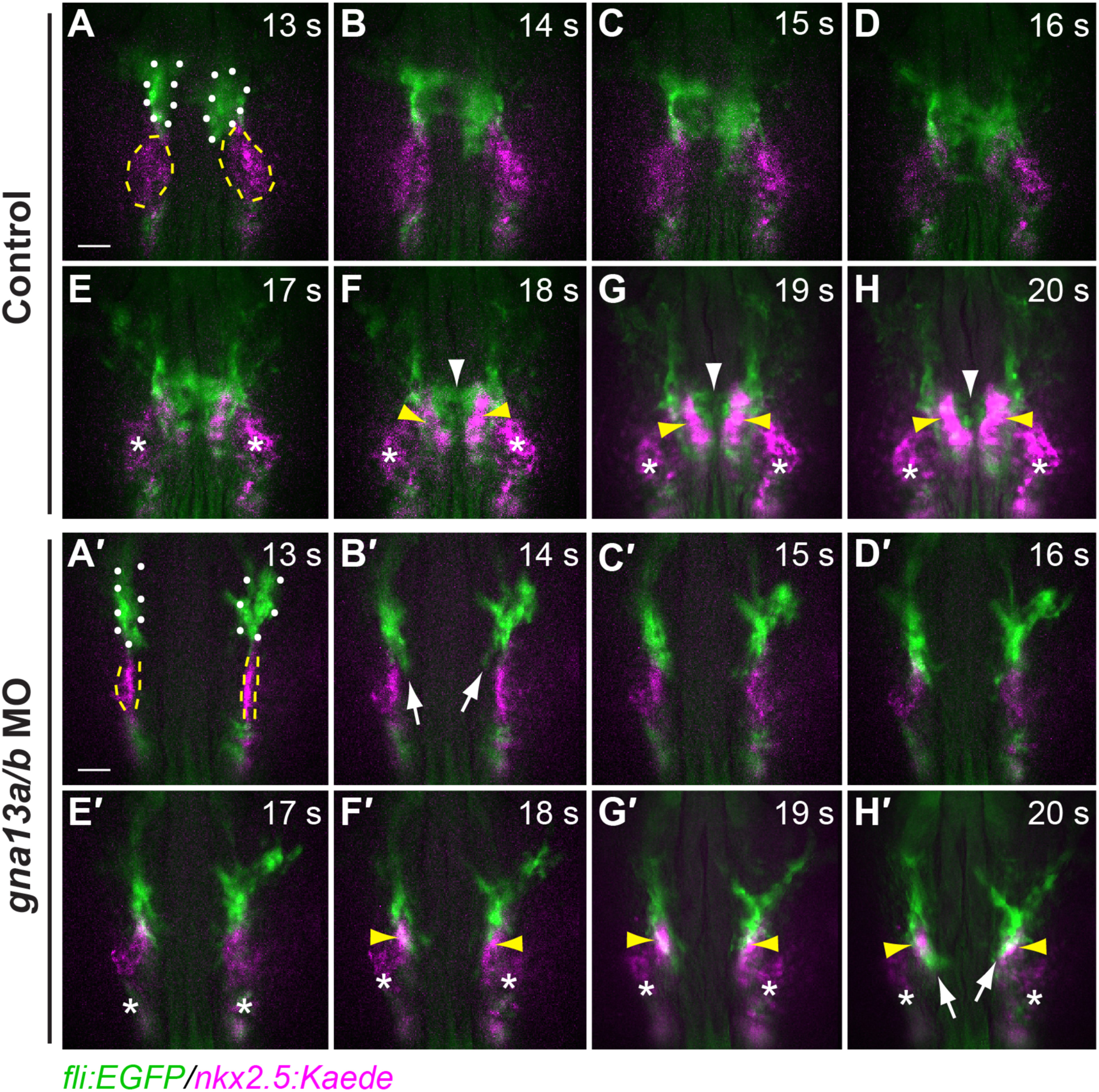
Fig. 2
In G?13-deficient embryos, endocardial precursors fail to migrate to the midline, instead moving toward the myocardial precursors. Epifluorescence time-lapse experiments were performed on control (A-H) and gna13a/b MO-injected (A?-H?) Tg(fli: EGFP/nkx2.5: Kaede) embryos (Supplementary movie 1) from 13 to 20 s Embryos were exposed to UV light at 10 s for 1 min to convert Kaede fluorescence from green to red (presented as magenta). Snapshots from movies showing the migration of endocardial (green, outlined by white dots in A, A?, white arrowheads in F-H) and myocardial (magenta, outlined by yellow dashed-lines in A, A?; yellow arrowheads in F?-H?) precursors at the stages indicated. White arrows: the leading region of migrating endocardial population. Asterisks: Non-myocardial cells labeled by Nkx2.5-Kaede. Dorsoanterior views. Scale bars: 100 �m.
Reprinted from Developmental Biology, 414, Xie, H., Ye, D., Sepich, D., Lin, F., S1pr2/G?13 signaling regulates the migration of endocardial precursors by controlling endoderm convergence, 228-43, Copyright (2016) with permission from Elsevier. Full text @ Dev. Biol.