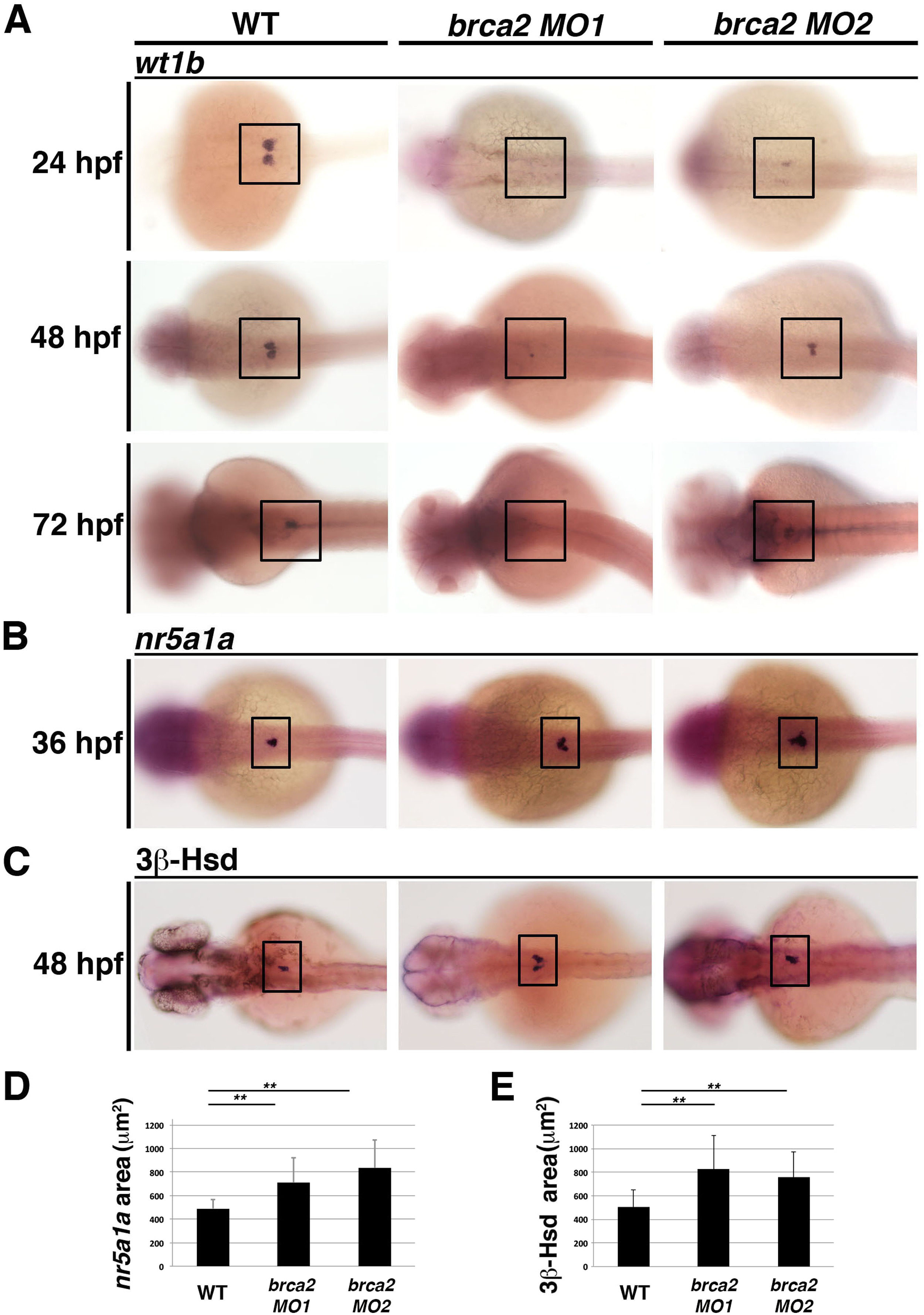
Fig. 7
Knockdown ofbrca2causes a reduction in expression of the podocyte markerwt1band an increase in the interrenal gland lineage. (A-C) At 24, 48 and 72 hpf, WT embryos display normal wt1b expression in podocytes, while microinjection of an MO targeting the brca2 start (MO1) or the splice acceptor of exon 21 (MO2) recapitulated the zep mutant phenotype, as observed by a reduction or complete abrogation of wt1b transcripts as assessed by WISH. Further, brca2 morphants had elevated (B) nr5a1a transcripts as assessed by WISH and (C) increased 3?-HSD chromogenic staining, which are quantified in (D,E), respectively. Asterisks (**) indicate p < 0.001 using student T-test. Embryos are shown in dorsal views, where black boxes demarcate the cervical region where podocytes and the interrenal gland develop.
Reprinted from Developmental Biology, 428(1), Kroeger, P.T., Drummond, B.E., Miceli, R., McKernan, M., Gerlach, G.F., Marra, A.N., Fox, A., McCampbell, K.K., Leshchiner, I., Rodriguez-Mari, A., BreMiller, R., Thummel, R., Davidson, A.J., Postlethwait, J., Goessling, W., Wingert, R.A., The zebrafish kidney mutant zeppelin reveals that brca2/fancd1 is essential for pronephros development, 148-163, Copyright (2017) with permission from Elsevier. Full text @ Dev. Biol.

