Fig. 6
Non-nuclear Sfpq Rescues Loss of Transcripts and Axonal Phenotype in the coma Mutant
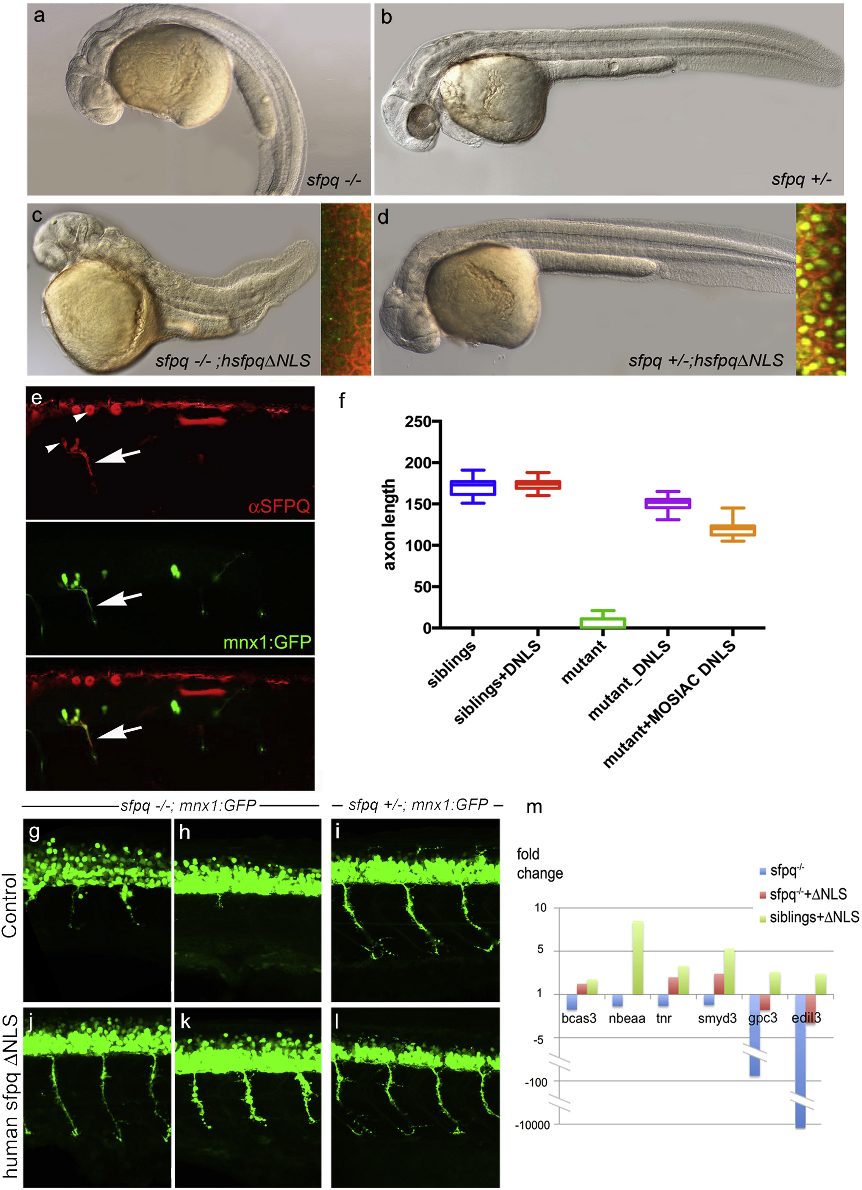
Lateral view of 30 hpf zebrafish (A?D) and spinal cord (G?L), anterior to the left.
(A?D) General morphology of sfpq?/? (A); sfpq+/? siblings (B); sfpq?/? injected with GFP-hsfpq?NLS (C) and sfpq+/? injected with GFP-hsfpq?NLS (D). Insets showing localization of the human GFP-tagged protein in early ss embryos, CAAX-Cherry in red.
(E) Whole-mount detection of SFPQ (red) in a 28 hpf sfpq?/? mutant, injected at one-cell stage with pmnx1:GFP and pCS2:hsfpq?NLS DNA, expressing these mosaically. Arrow shows motor axon. Note the negative nuclei (arrowhead).
(F) Measurement of ventral motor axon length in siblings and homozygous mutant uninjected or injected with RNA (DNLS) or DNA (MOSAIC DNLS) coding for hSFPQ?NLS. For the controls and RNA injections, the ventral motor axons were measured in five embryos over five-somite length upstream of the cloaca. For mosaic DNA-injected sample, measures were done only in seven homozygous mutants, in the same trunk area for the rare SFPQ+ neurons.
(G?L) Lateral view, anterior to the left of spinal cords, showing rescue of the axonal defect both in the anterior (G and J) and posterior (H and K) trunk of sfpq?/?;Tg (mnx1:GFP) by injection of the ?NLS human SFPQ. Sibling axons are unaffected by the injection (I and L).
(M) qPCR results for six out of ten DES transcripts from 32 hpf siblings and sfpq?/? mutant embryos uninjected or injected with human ?NLS sfpq RNA. Bars show expression fold changes compared to the expression level of the transcript in uninjected sibling embryos taken as reference. The four transcripts not plotted did not show any improvement after ?NLS rescue.

