
|
Fig. S4
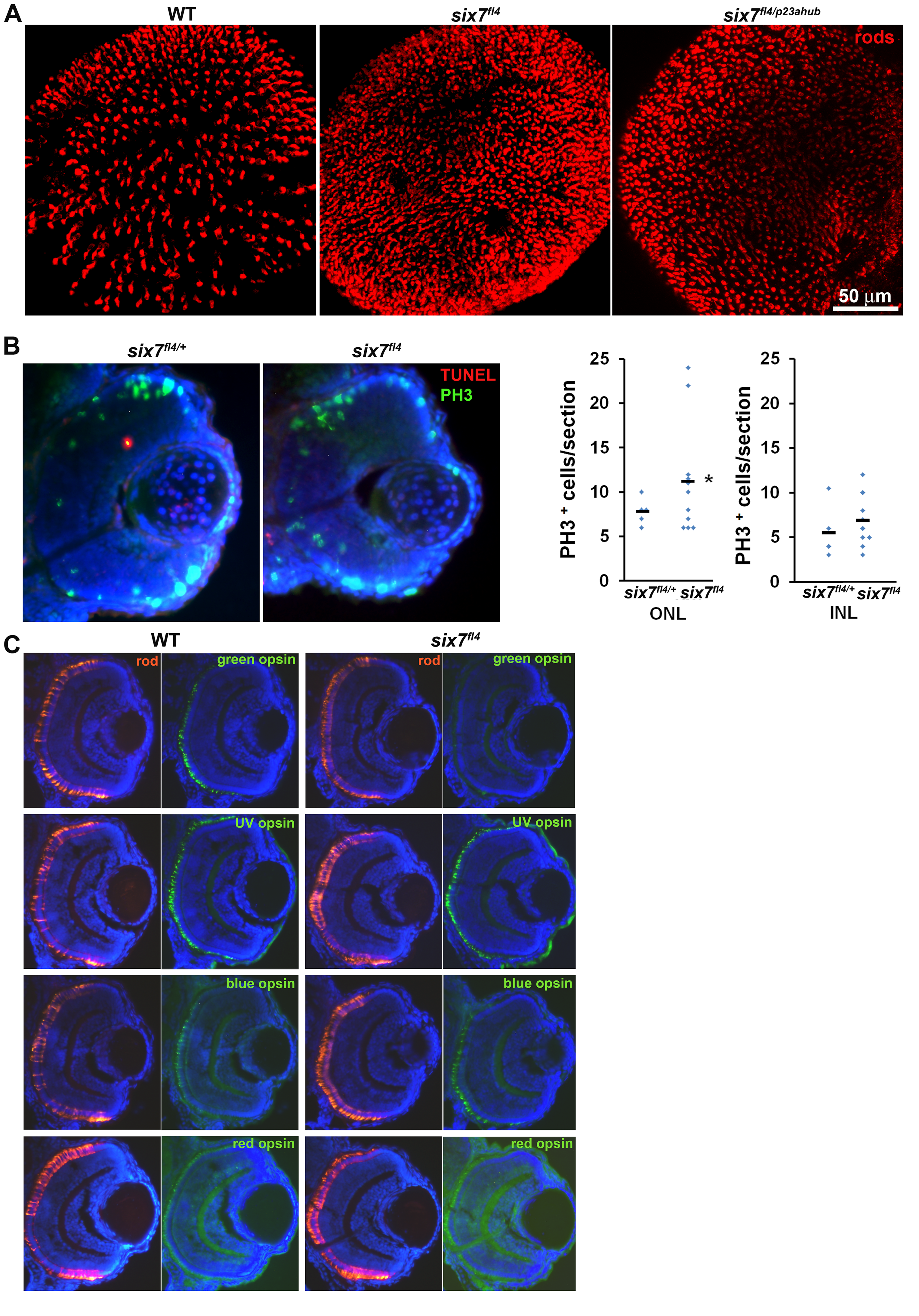
Levels of mitosis are altered in six7fl4 at 56 hpf.
(A) Confocal immunofluorescent images labeled for rods (4C12, red) from WT, six7fl4 and six7fl4/p23ahub retinas at 4 dpf. (B) Retinal cryosections from carrier animals (six7fl4/+, n = 5, 1?2 sections/retina) and six7 (n = 11) embryos at 56 hpf co-labeled for TUNEL (red) and PH3 (green), nuclei counterstained with DAPI. No differences in TUNEL labeling were detected. Graphs showing the number of PH3+ cells by section (excluding CMZ). Number of PH3+ cells is significantly greater in ONL of six7fl4 mutants at 56 hpf. Un-paired Student t test with Welch?s correction, *p<0.05). (C) Retinal cryosections from WT and six7fl4 embryos at 4 dpf immunolabeled for rods (4C12, red) and the green-, UV-, blue- and red-sensitive opsins (green). Nuclei were counterstained with DAPI (blue). Dorsal is up. Depleted green-sensitive opsin expression is noticeable in six7fl4, but other opsins appear unaltered.