
|
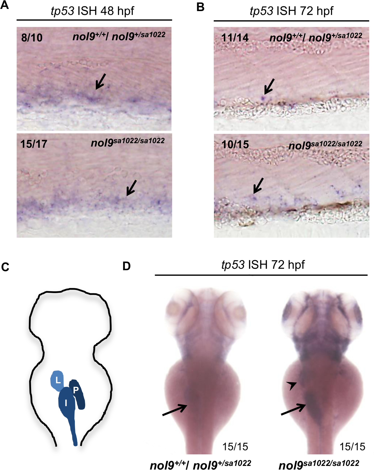
Fig. 8
nol9sa1022/sa1022 embryos display tissue-specific upregulation of tp53.
Representative images of embryos stained by whole-mount in situ hybridization against tp53. (A) At 48 hpf, similar levels of tp53 signal (arrow) was detected in the CHT of nol9sa1022/sa1022 embryos and their wild-type siblings. (B) At 72 hpf, nol9sa1022/sa1022 mutant embryos were characterized by more tp53 signal in the CHT than their wt siblings. Mann-Whitney U test, p<0.05. (A-B) All embryos are oriented with anterior to the left and dorsal to the top. (C) Schematic representation of digestive organs in a wild-type 72 hpf zebrafish larva. I?intestine, L- liver, P- pancreas. (D) At 72 hpf, nol9sa1022/sa1022 mutant embryos display strong tp53 signal in the liver (arrowhead) and intestine (arrow), compared to weak signal in the intestine of wild-type siblings. (C-D) Dorsal view anterior up.