
Fig. 1
|
Fig. 1
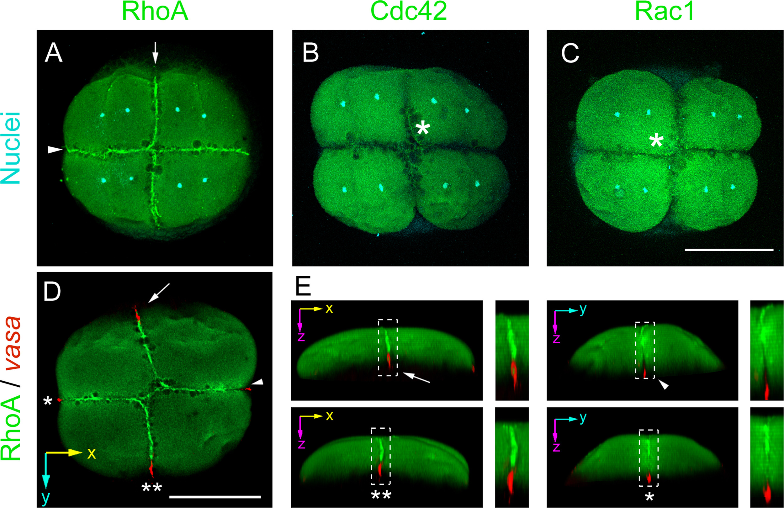
Rho GTPases immunofluorescense pattern during germ plasm localization at 8-cell stage zebrafish embryos. (A) RhoA immunolocalization. Arrow indicates first cleavage and arrowhead second cleavage furrows that are positive for RhoA (n=31 of 6 independent experiments). (B) Cdc42, immunolocalization (n=8 of 2 independent experiments). (C) Rac1 immunolocalization (n=7 of 2 independent experiments). Asterisks indicate cleavage furrow. (A, B and C) Nuclei were stained with DAPI and are shown in cyan. (D) RhoA (green), immunolocalization and vasa mRNA (red), localization by fluorescent in situ hybridization (n=9 in 2 independent experiments). (E) 3D reconstruction models of the image shown in D, rotated to highlight the regions indicated by the arrow, the single asterisk, the arrowhead and the double asterisks. The inserts indicated by the discontinuous lined squares are magnified and shown on the right side of each 3D model. Scale bar 250 μm.
Reprinted from Developmental Biology, 421(1), Miranda-Rodríguez, J.R., Salas-Vidal, E., Lomelí, H., Zurita, M., Schnabel, D., RhoA/ROCK pathway activity is essential for the correct localization of the germ plasm mRNAs in zebrafish embryos, 27-42, Copyright (2017) with permission from Elsevier. Full text @ Dev. Biol.