
|
Fig. 3
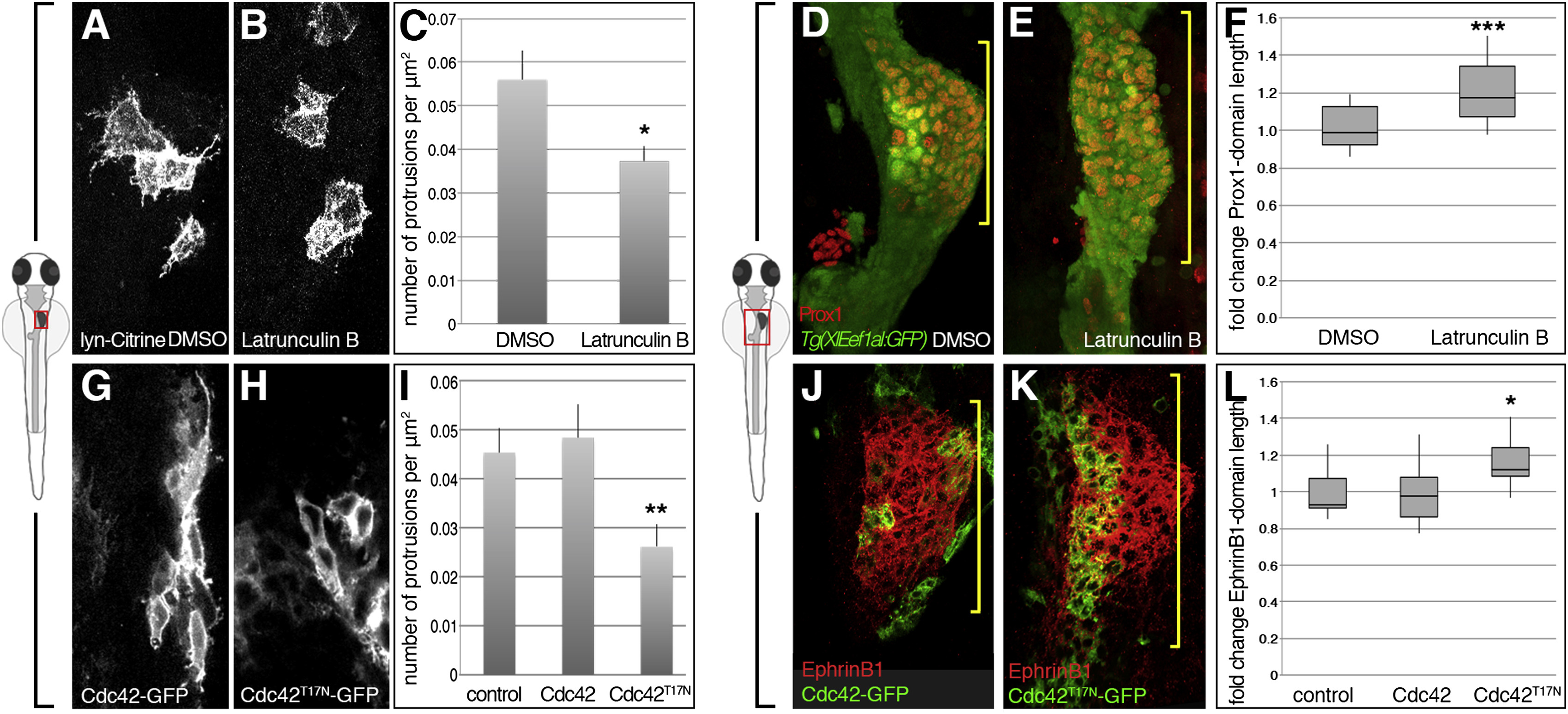
Compromised Protrusion Formation Correlates with Liver Budding Defects
(A-F) Latrunculin B (0.1 ?g/ml) treatment during liver budding (26-32 hpf) leads to significantly less hepatoblast protrusions (A-C; DMSO clones, n = 7; Lat B clones, n = 9, N = 1), and a significantly longer Prox1 domain (bracket) (D?F; DMSO, n = 23; Lat B, n = 13, N = 2).
(G-L) Hepatoblasts expressing Cdc42T17N-GFP during liver budding (26?32 hpf) form significantly less protrusions compared to controls and Cdc42-GFP (G-I; control clones, n = 10; Cdc42-GFP clones, n = 16; Cdc42T17N-GFP clones, n = 18, N = 1) and a significantly longer EphrinB1 domain (bracket) (J?L; control, n = 7; Cdc42-GFP, n = 18; Cdc42T17N-GFP, n = 16, N = 2). N indicates the number of experiments.
?p < 0.05, ??p < 0.01, ???p < 0.001.
Reprinted from Developmental Cell, 39, Cayuso, J., Dzementsei, A., Fischer, J.C., Karemore, G., Caviglia, S., Bartholdson, J., Wright, G.J., Ober, E.A., EphrinB1/EphB3b Coordinate Bidirectional Epithelial-Mesenchymal Interactions Controlling Liver Morphogenesis and Laterality, 316-328, Copyright (2016) with permission from Elsevier. Full text @ Dev. Cell