Fig. S2
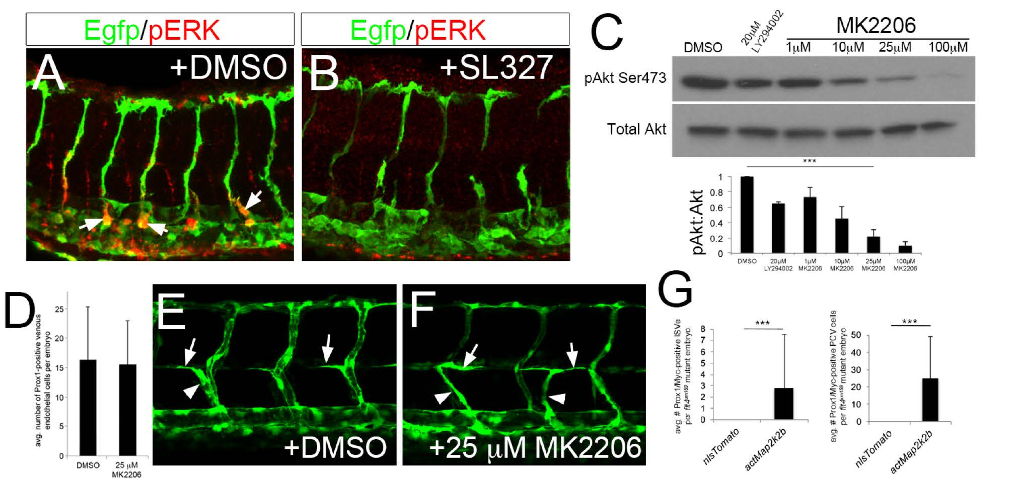
Akt is dispensable for early lymphatic development. (A, B) Wild type Tg(fli1a:egfp)y1 at 36 hpf immuonstained for pERK (red) and EGFP (Green). Embryos were treated with (A) DMSO or (B) 15 ?M SL327 starting at 28 hpf. (C) pAkt and total Akt levels in whole embryo lysates treated with indicate compound at indicated concentration. Graph shows quantification of Akt phosphorylation intensity from Western normalized to total Akt levels. ***p<0.001, error barsąS. D. 25 ?M MK2206 significantly reduce pAkt levels by 80% and was used for subsequent studies. (D) Quantification of Prox1-positive endothelial cells in embryos treated with DMSO or 25 ?M MK2206 between 28 and 38hpf. (E, F) Confocal microscopy of live Tg(fli1a:egfp)y1 at 49 hpf treated with (E) DMSO or (F) 25 ?M MK2206 from 20 hpf. Parachordal cells (arrows) and lymphatic sprouts (arrowheads) are apparent in both DMSO and MK2206-treated embryos. (G) Quantification of lymphatic sprouts and Prox1/Mycpositive posterior cardinal vein (PCV) endothelial cells in flt4C562? mutant embryos injected with indicated transgene.

