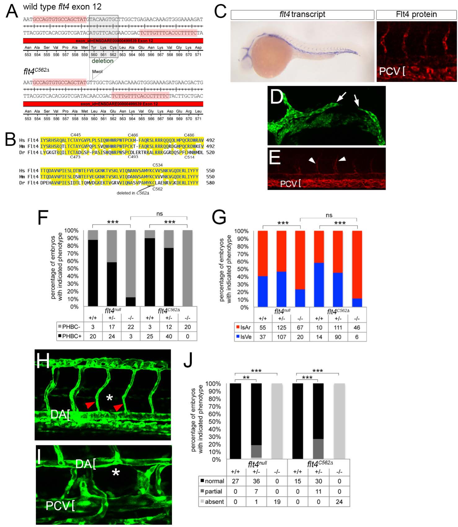
Fig. S1
Generation and characterization of flt4C562?. (A) TALEN target sequences (highlighted in pink) in flt4 exon 12 flanking Cysteine 562. Boxed sequence indicates amino acids deleted in flt4C562? mutants. The nine nucleotide deletion creates an MwoI site used for genotyping. (B) Alignment of region of Flt4 extracellular domains from human (Hs), mouse (Mm), and zebrafish (Dr) containing cysteines implicated in disulfide linkage between cleaved extracellular domain and remaining Flt4 protein. Position of amino acids deleted in flt4C562? is indicated. (C) Left, whole mount in situ hybridization using a digoxigenin labeled antisense flt4 riboprobe on flt4C562? mutant embryos at 25 hpf. Right, whole mount immunostaining using a polyclonal antibody against zebrafish Flt4 at 30 hpf; flt4 transcript and Flt4 protein are detectable in flt4C562? mutant embryos. (D) Confocal image of flt4C562? mutant Tg(fli1a:egfp)y1 embryo at 26 hpf; incompletely formed primordial hindbrain channel is indicated by white arrows. (E) Tie2 immunostained flt4C562? mutant embryo showing ISVe loss. White arrowheads denote weak staining in ISAs (F) Percentage of embryos with delayed PHBC formation of indicated genotype. (G) Percentage of ISA and ISVe connections in embryos of indicated genotype at 72 hpf. (F, G) Data from flt4null are same as those shown in main figures. (H) Tg(fli1a:egfp)y1 embryo mutant for flt4C562? at 48 hpf showing lack of parachordal cells (PACs; normal position indicated by asterisk); ISAs indicated by red arrowheads, dorsal aorta denoted by bracket. (I) Loss of thoracic duct in Tg(fli1a:egfp)y1 embryo mutant for flt4C562? at 5 dpf. Normal position of thoracic duct denoted by asterisk; dorsal aorta (DA) and posterior cardinal vein (PCV) are indicated by brackets. (J) Percentage of embryos of indicated genotype with normal, partial, or absent thoracic duct at 5 dpf. Data from flt4null are the same as those shown in Figure 2

