Fig. 4
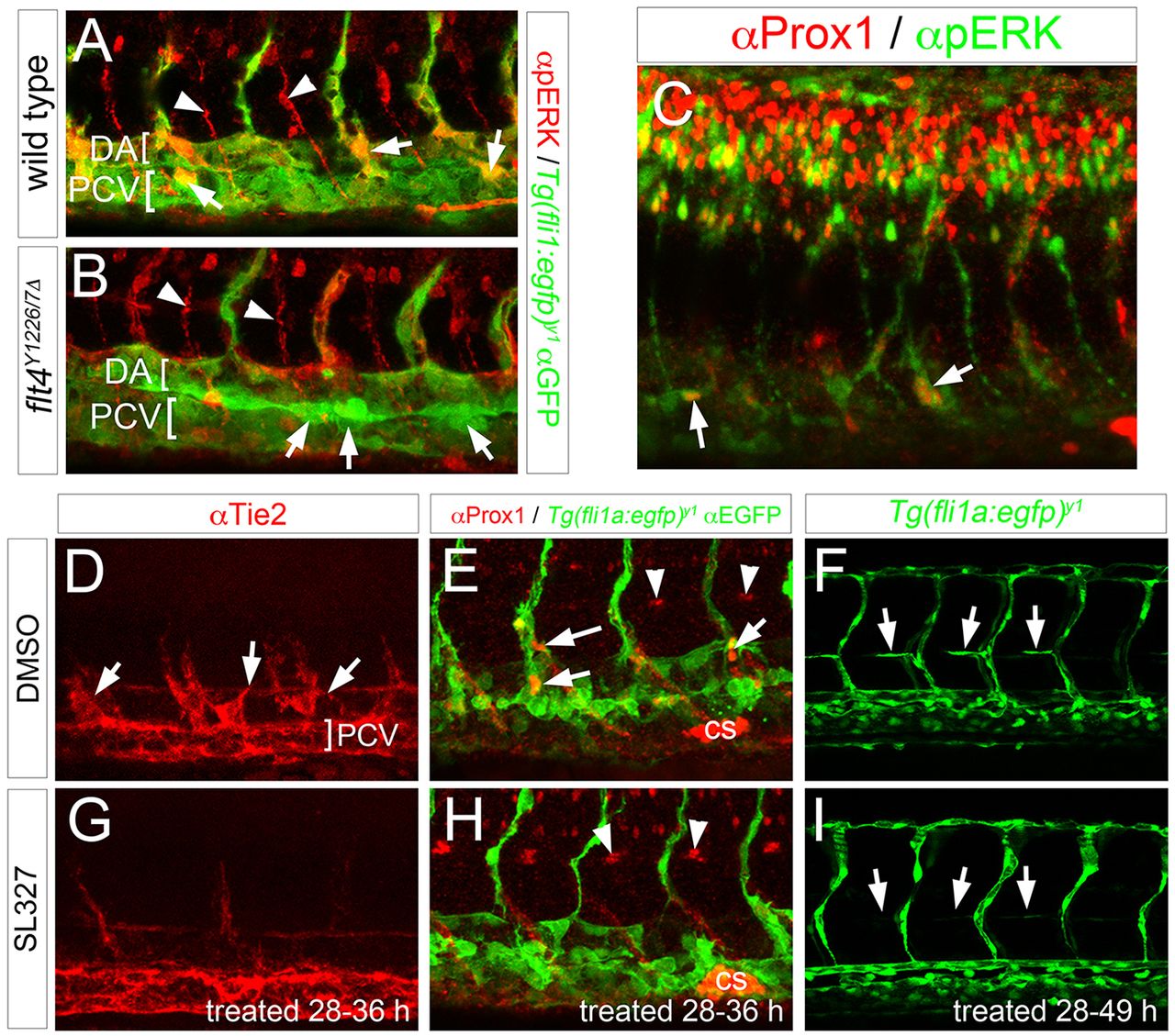
ERK signaling is essential for trunk lymphatic morphogenesis and differentiation. (A-I) Confocal images; lateral views, anterior to the left, dorsal is up. (A,B) pERK (red) and GFP (green) immunostaining in Tg(fli1a:egfp)y1 embryos of the indicated genotype at 34?hpf. Arrows denote pERK-positive lympho-venous sprouts, arrowheads indicate pERK staining in motor neurons. Brackets indicate DA and PCV. (C) Prox1 (red) and pERK (green) immunostaining in a wild-type embryo at 38?hpf. Arrows denote endothelial cells that are positive for Prox1 and pERK in lymphatic sprouts or dorsal wall of PCV. (D-I) Embryos treated with DMSO (D-F) or 15?ÁM SL327 (G-I). Treatment time is indicated. (D,G) Tie2 immunostaining; arrows denote lympho-venous sprouts, PCV indicated by bracket. (E,H) Prox1 (red) and GFP (green) immunostaining of Tg(fli1a:egfp)y1 embryos at 36?hpf; arrows denote Prox1-positive lymphatic sprouts, arrowheads indicate muscle pioneers and Corpuscle of Stannius (cs) is indicated. (F,I) Live Tg(fli1a:egfp)y1 embryos at 49?hpf. Arrows indicate parachordal cells, or absence thereof.

