
|
Fig. 4
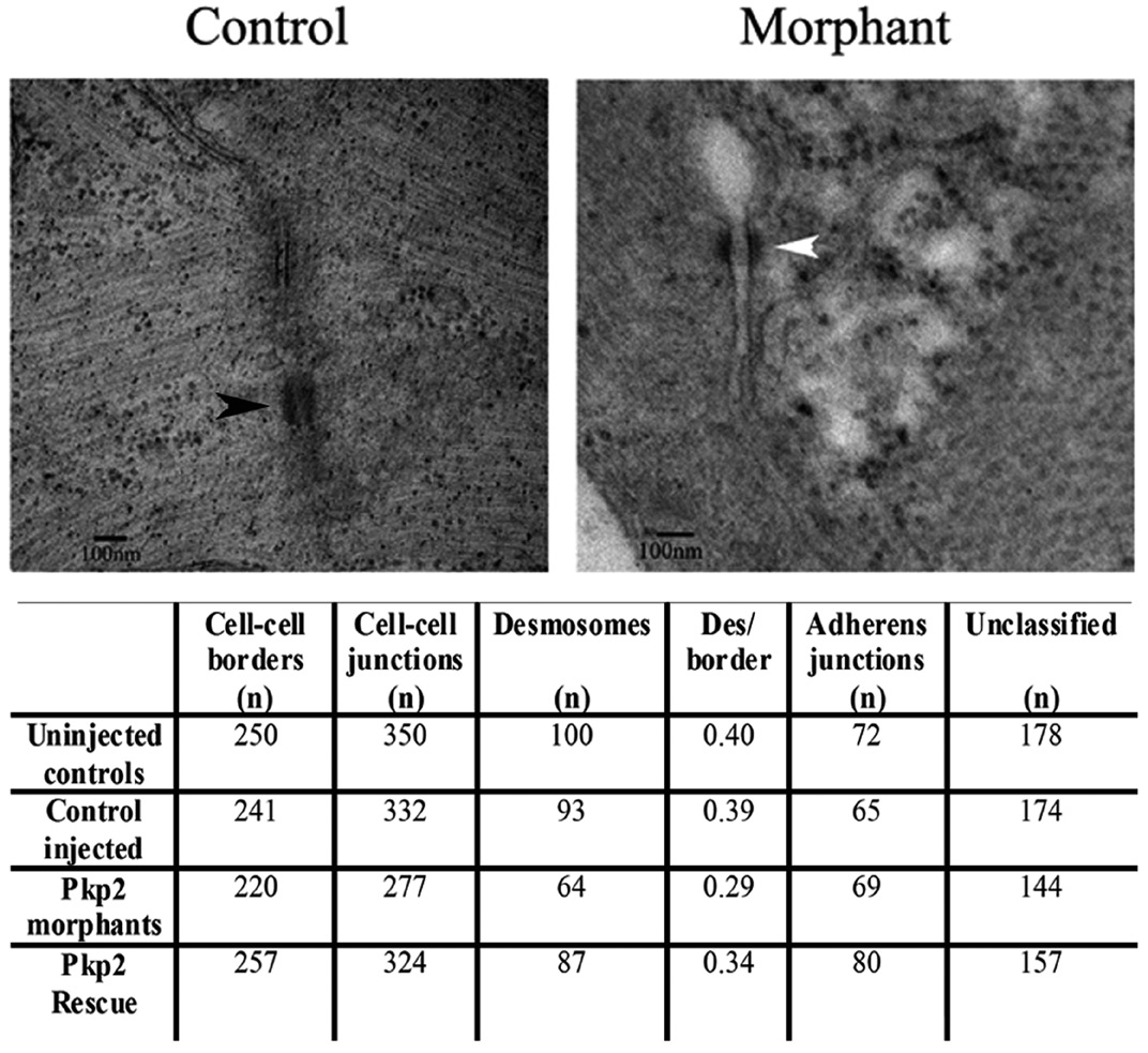
The ultrastructure of adhesion junctions in plakophilin 2 morpholino injected embryo hearts. Electron microscopy of the intercalated discs of zebrafish embryo hearts at 72 hpf. (A) In control morpholino injected embryos, intercellular junction elements (black arrowhead) were clearly evident. (B) In plakophilin 2 morphant embryos, junctions lacked these intercellular elements (white arrowhead) and the gap between cells was larger. (C) The table details the cell–cell border numbers, the number of adhesion junctions, and the number of each type of junction in control, morphant and rescued embryos. The number of desmosomes per border was decreased in plakophilin 2 morphants compared to controls, with an increase towards normal levels in the rescued embryos. Unclassified indicates adhesion junctions that could not be definitively classed as adherens junctions or desmosomes. N=3 embryos per sample. Scale bar is 100 nm.