Fig. 4
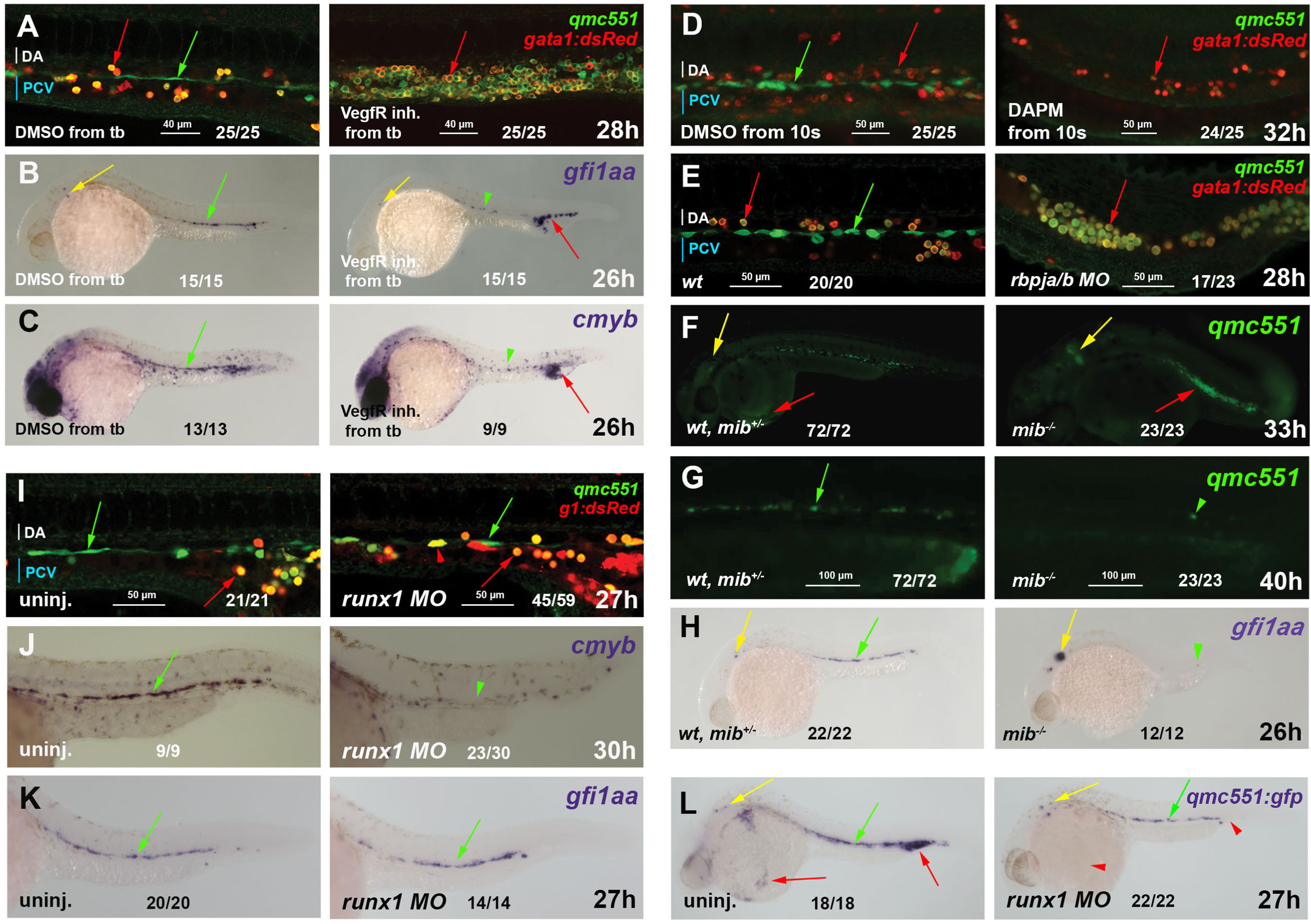
Gfi1aa expression in haemogenic endothelial cells is induced downstream of VegfA and Notch signaling, but independent of Runx1. Fixed qmc551;gata1:dsRed double transgenic embryos after GFP/dsRed immunohistochemistry are shown in (A, D-E, I). Fixed wt, qmc551 and qmc551;gata1:dsRed double transgenic embryos stained by WISH are shown in (B-C, H, K), (L) and (J), respectively. Live qmc551 embryos that were wt, heterozygous or homozygous mib carriers were imaged in (F-G). Confocal images of optical sagittal sections through the DA are 1.6 and 0.995 Ám (A), 6.5 and 6.6 Ám (D), 1.2 Ám (E) and 2.7 Ám (I) thick. A confocal maximum intensity projection of a 37 Ám optical slice is shown in (G). Embryos were treated with DMSO, the VegfR inhibitors 676,475 (A) and SU5416 (B,C) or DAPM (D) from tailbud stage (10 hpf). Rbpja/b (E) and runx1 (I-L) morpholinos were injected at 2?4 cell stage. PrRBCs, HECs and inner ear hair cells are labeled with red, green and yellow arrows, respectively. Arrowheads mark reduced or absent staining. Fractions x/y give the number of embryos, x, with depicted phenotype out of all embryos analyzed, y. Embryos are shown with anterior left and dorsal up.
Reprinted from Developmental Biology, 417(1), Thambyrajah, R., Ucanok, D., Jalali, M., Hough, Y., Wilkinson, R.N., McMahon, K., Moore, C., Gering, M., A gene trap transposon eliminates haematopoietic expression of zebrafish Gfi1aa, but does not interfere with haematopoiesis, 25-39, Copyright (2016) with permission from Elsevier. Full text @ Dev. Biol.

