
|
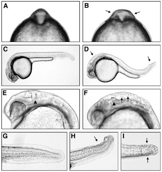
Fig. 1
pac mutants exhibit abnormal brain and tail morphogenesis. (A,C,E,G) Wild-type siblings, (B,D,F,H,I) pacfr7 mutants. (A,B) Eight-somite stage, frontal view, arrows in B indicate broadened neural tube at midbrain-hindbrain level. (C-I) 24 hpf. (C,D) lateral overview; arrows in D indicate sites of phenotypic defects in the midbrain-hindbrain region and tip of tail. (E,F) Magnification of head region; arrowheads in E,F indicates the position of the midbrain-hindbrain boundary, arrows in F indicate loose cell aggregates in ventricle. (G-I) Magnification of tip of tail; lateral view (G,H) and dorsal view (I); arrows in H,I indicate region with split dorsal fin.