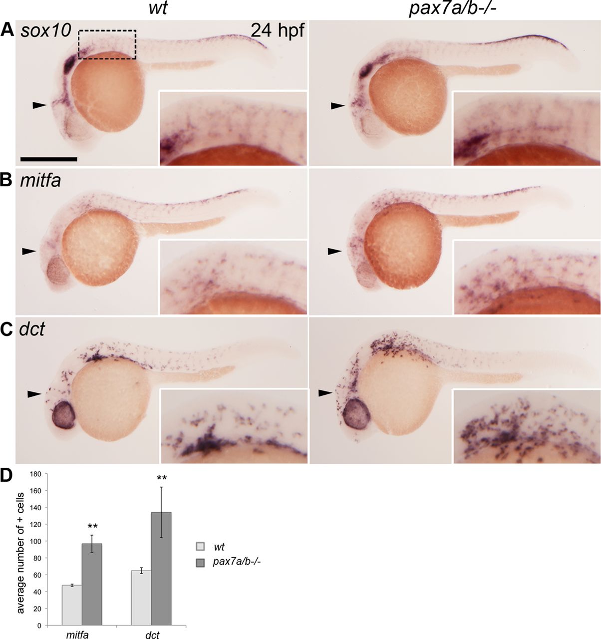
Fig. 3 The pax7a/pax7b double mutants have an increased number of melanoblasts. Whole-mount in situ hybridization on wt siblings and pax7a/pax7b double-mutant zebrafish embryo at 24 hpf showing the expression of (A) sox10 (n > 50 for siblings and n = 7 for pax7a/pax7b double mutants), (B) mitfa (n = 5 and 5), and (C) dct (n = 7 and 3). (D) Average number of mitfa+ and dct+ cells in the region anterior to the first somite on one side of wt siblings and pax7a/pax7b double-mutant embryos at 24 hpf; positive cells in the eye were excluded. Student?s t test was used to calculate significance; **p < 0.01. Error bars indicate SEM. Box indicates area of enlargement visualized in insets. Arrowheads indicate head region where changes in expression can be detected. Scale bar, 200 μm.
Image
Figure Caption
Figure Data
Acknowledgments
This image is the copyrighted work of the attributed author or publisher, and
ZFIN has permission only to display this image to its users.
Additional permissions should be obtained from the applicable author or publisher of the image.
Full text @ Mol. Biol. Cell

