Fig. 1
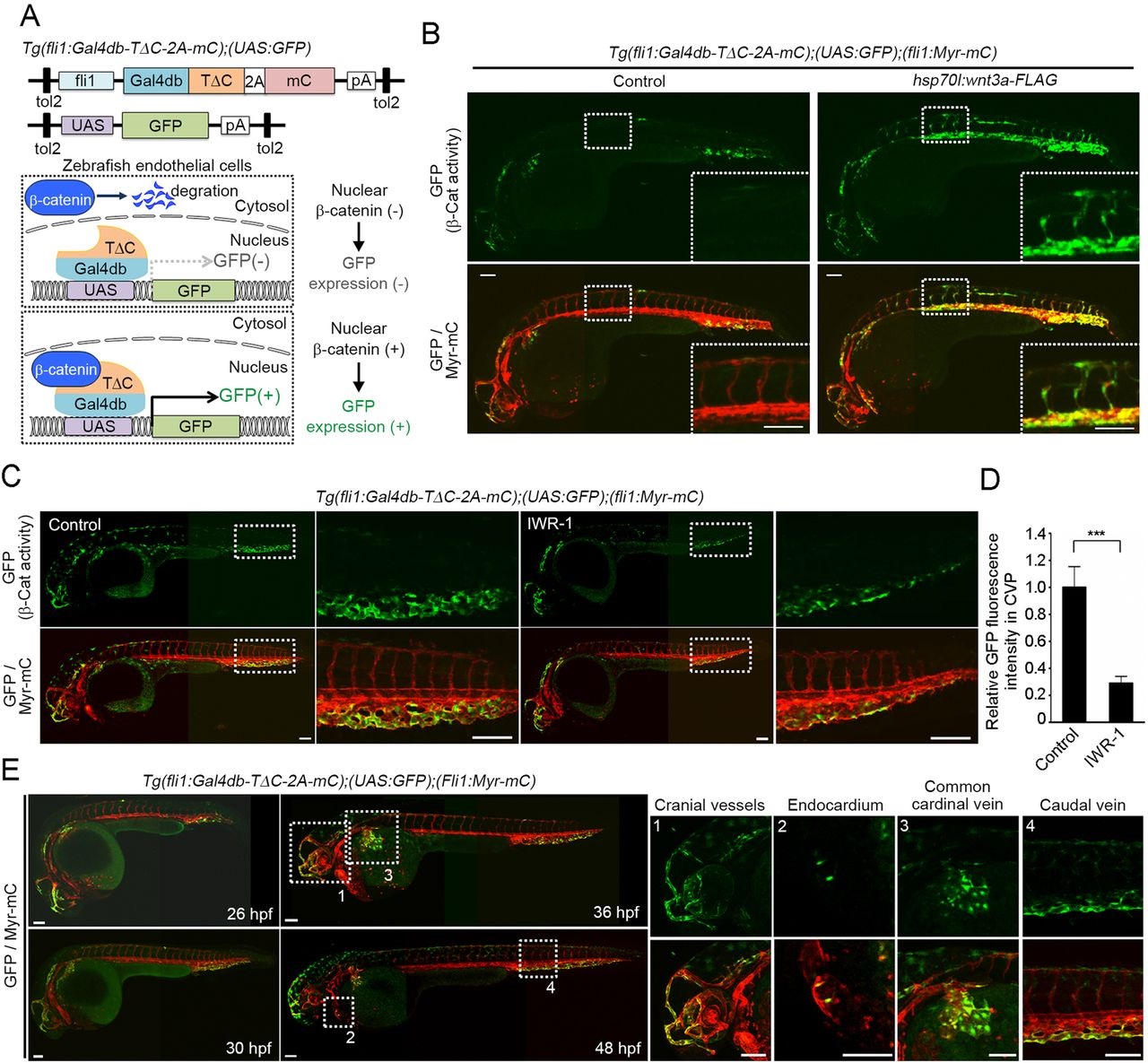
Generation of an endothelial cell-specific β-catenin reporter zebrafish line. (A) Schematic representation of the endothelial cell (EC)-specific β-catenin reporter system. (Top) The constructs used to generate the EC-specific β-catenin reporter zebrafish line; (bottom) how the system works. In the absence of upstream signaling, β-catenin undergoes proteasomal degradation in the cytoplasm. Upon induction of the signaling that promotes β-catenin stabilization, β-catenin translocates to the nucleus and binds Gal4db-TΔC, thereby inducing GFP expression. Thus, this fluorescence reflects the transcriptional activity of β-catenin in ECs. Gal4db, DNA-binding domain of Gal4; TΔC, β-catenin-binding domain of Tcf4; 2A, 2A peptide sequence; mC, mCherry; pA, polyadenylation signal; UAS, upstream activation sequence. (B) 3D-rendered confocal stack fluorescence images of 32hpf Tg(fli1:Gal4db-TΔC-2A-mC);(UAS:GFP);(fli1:Myr-mC) embryos injected without (control) or with hsp70l:wnt3a-FLAG plasmid and heat shocked at 22hpf for 1h. (Top) GFP images (β-catenin activity); (bottom) merged images (GFP/Myr-mC) of GFP (green) and mCherry (red). The boxed areas are enlarged in the insets. All confocal fluorescence images are lateral views with anterior to the left unless otherwise described. Myr-mC, myristoylation signal-tagged mCherry. (C) Confocal images of embryos treated with vehicle (control) or IWR-1, an axin-stabilizing compound, from 15-36hpf, as in B. The boxed areas are enlarged to the right. (D) Fluorescence intensity of GFP in the caudal vein plexus (CVP), as observed in C, relative to that observed in vehicle-treated embryos. Data are meanħs.e.m. Control, n=7; IWR-1, n=11. ***P<0.001. (E) Merged fluorescence images (GFP/Myr-mC) of GFP (green) and mCherry (red) in Tg(fli1:Gal4db-TΔC-2A-mC);(UAS:GFP);(fli1:Myr-mC) embryos at 26, 30, 36 and 48hpf. The boxed areas labeled 1-4 are enlarged to the right, showing (top) GFP images (β-catenin activity) and (bottom) merge of GFP (green) and mCherry (red) (GFP/Myr-mC). Note that green signal that does not overlap with mCherry fluorescence is background autofluorescence of the zebrafish embryos. Panels B, C and E are composites of two or three images, since it was not possible to capture the whole animal at sufficiently high resolution in a single field of view. Scale bars: 100µm.

