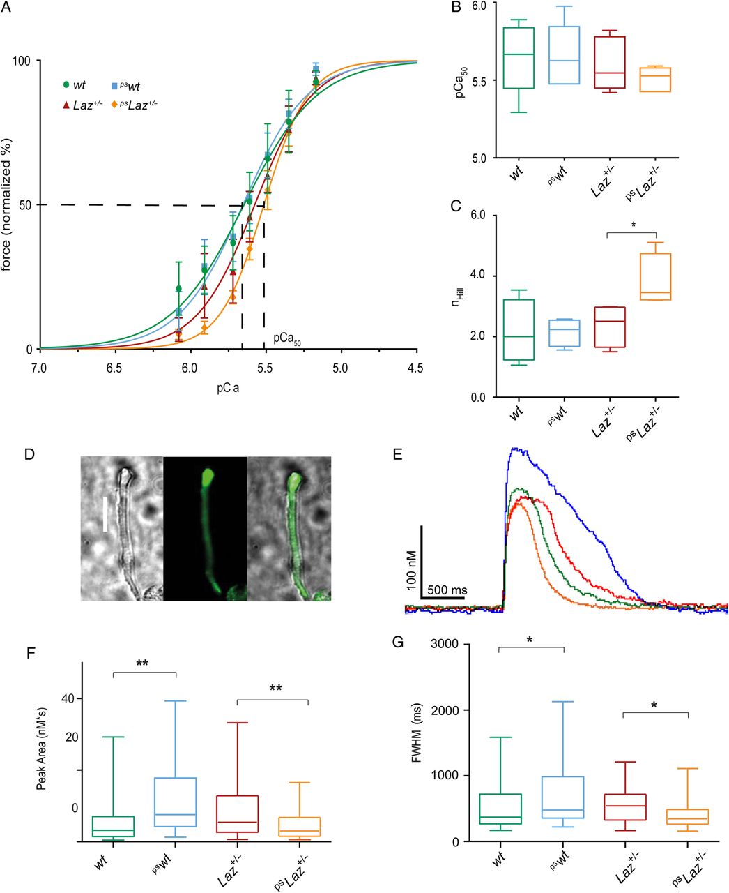
Fig. 5
Laz affects acto-myosin cooperativity and impairs calcium handling.
(A) pCa-force relationship, normalized to maximal force (at pCa 4.5). The sigmoidal curves were further analysed for pCa50 and nHill. (B) Quantification of pCa50 from the pCa-force relationship revealed no significant differences. (C) nHill was significantly increased for pslaz+/- compared with wt, pswt, and laz+/- (P < 0.05; N = 5). (D) Intracellular Ca2+-transients were analysed in isolated ventricular cardiomyocytes loaded with Fluo4-AM. Representative brightfield (left) and corresponding fluorescent (middle) and merged (right) images of a single ventricular adult zebrafish cardiomyocyte (scale bar 20 Ám). (E) Representative Ca2+-transient traces recorded from wt, pswt, laz+/-, and pslaz+/-. (F) Peak area and (G) full-width half-maximum (FWHM) obtained from the analyses of Ca2+-transients. Data in (F) and (G) were statistically analysed by using ANOVA with Bonferonni post hoc pairwise multiple comparisons *P < 0.05; **P < 0.01; in A-C: N = 5; in E-G: n = 90 cells.

