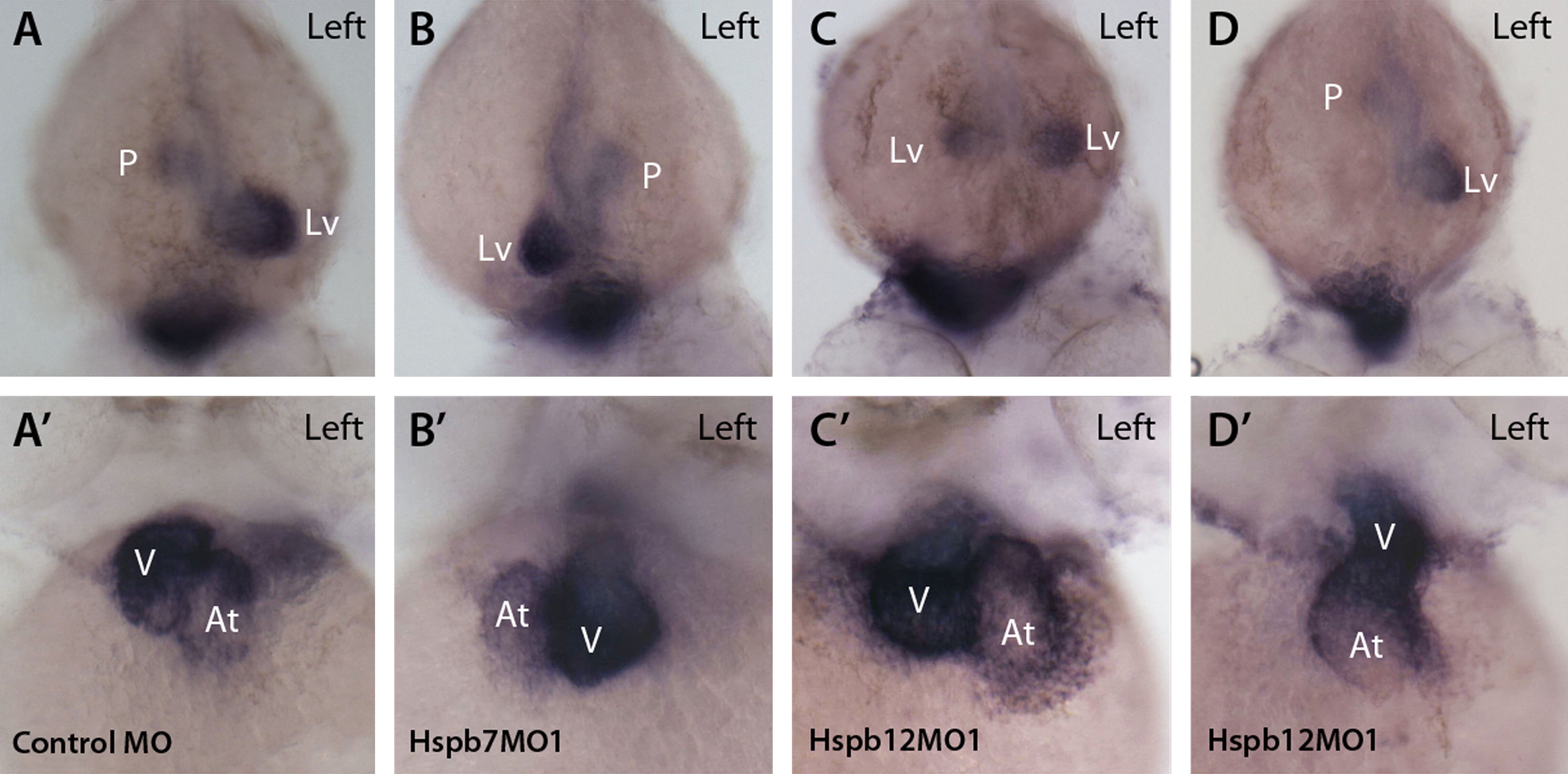
Fig. 6
Concordance of asymmetry between heart and gut in hspb7 and hspb12 knockdown embryos. (A-D) Dorsal view, rostral toward bottom, left side as indicated in (A). Expression of foxa3 is strong in liver (Lv) and weaker in gut and pancreatic bud (P) at 48 hpf. (A′-D′) myl7 (cmlc2) is strongly expressed in ventricle (V) and more weakly in atrium (At) at 48 hpf. Anterior view, left side as indicated in (A). (A, A′) Control MO-injected embryo demonstrated the normal position of atrium to the left of the embryo′s midline and of the ventricle on the right (dextral loop). foxa3 is concentrated in the normal, left-sided liver and weaker in the pancreatic bud in control MO-injected embryo. (B, B′) hspb7 morphant with right-sided liver and ventricle. (C, C′) hspb12MO1-injected embryo with bilateral liver but normal position of the atrium and ventricle. (D, D′) hspb12MO1-injected embryo with left-sided liver and a heart that failed to jog or loop (midline heart).
Reprinted from Developmental Biology, 384(2), Lahvic, J.L., Ji, Y., Marin, P., Zuflacht, J.P., Springel, M.W., Wosen, J.E., Davis, L., Hutson, L.D., Amack, J.D., and Marvin, M.J., Small heat shock proteins are necessary for heart migration and laterality determination in zebrafish, 166-180, Copyright (2013) with permission from Elsevier. Full text @ Dev. Biol.

