
Fig. S1
|
Fig. S1
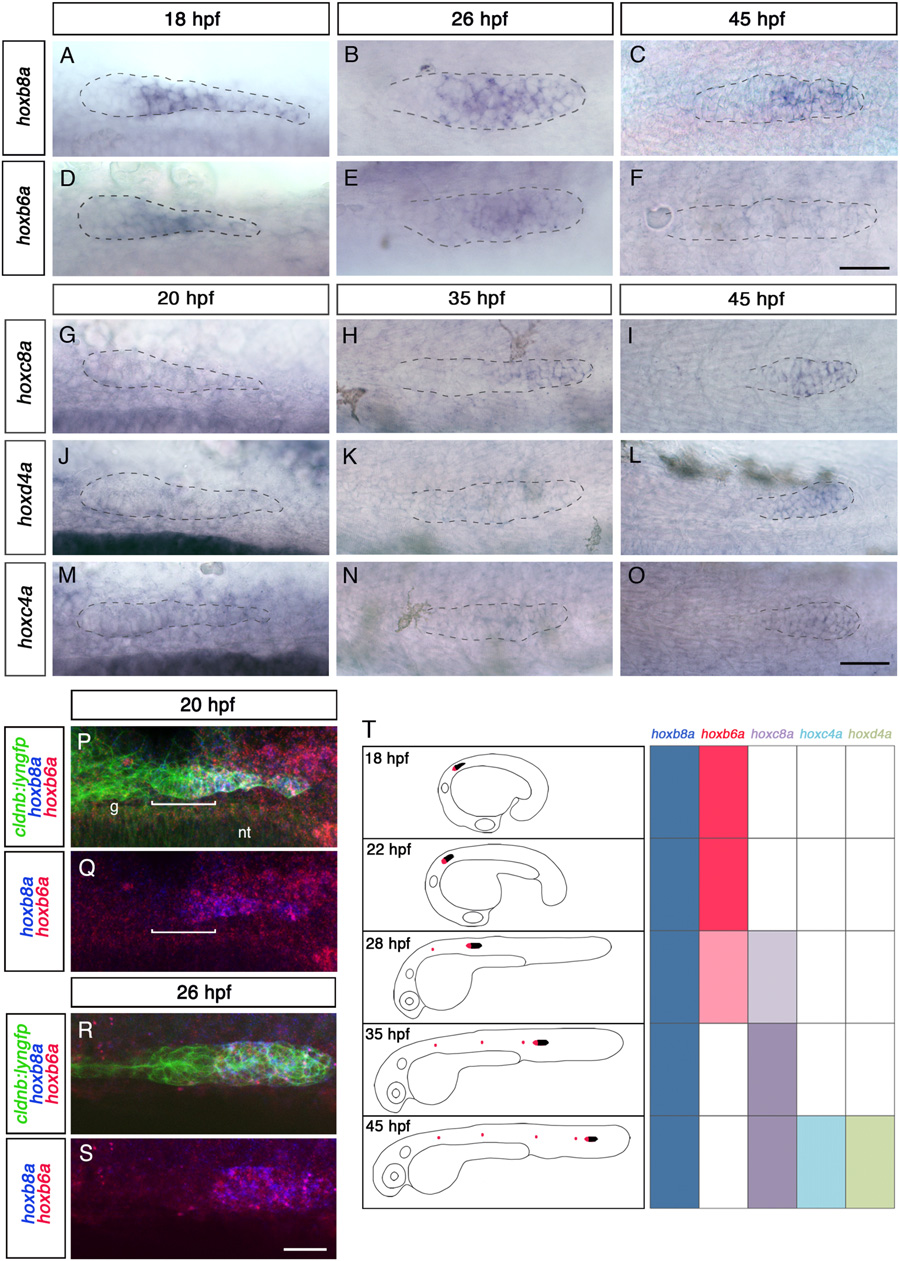
Expression pattern of hox genes in the posterior lateral line primordium. (A-O) Expression of hoxb8a, hoxb6a, hoxc8a, hoxd4a, and hoxc4a detected by in situ hybridization (ISH) at 18 or 20 h postfertilization (hpf), 26 hpf, 35 hpf, and 45 hpf. Dorsal view at 18 and 20 hpf and lateral view at later stages. The primordium is outlined with dotted line. (P-S) Fluorescent detection of hoxb8a (blue) and hoxb6a (red) mRNA in cldnb:lyngfp (green) embryos at 20 (P and Q, dorsal view) and 26 hpf (R and S, lateral view). White bracket in P and Q indicates the width of the first proneuromast (L1). (T) Summary of spatiotemporal expresssion of hox genes during migration of the primordium. (Left) The developmental stages and extent of the primordium migration, with the hox expression domain in the leading zone indicated in black. (Right) Table shows when different hox genes are expressed; hoxc6a expression pattern is similar to that of hoxc8a. g, Posterior lateral line sensory ganglion; nt, neural tube. (Scale bars: 25 µm.)