
|
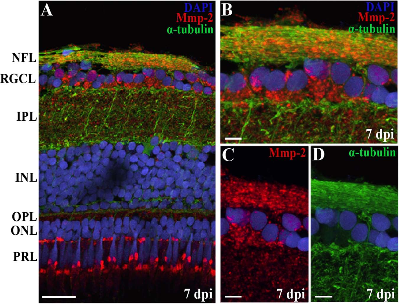
Fig. 6
Overlap of α-tubulin and Mmp-2 expression on retinal sections localizes Mmp-2 in/on axons of the NFL. A-D: A double immunostaining for α-tubulin and Mmp-2 on retinal sections shows a similar expression of both proteins in the NFL and thus indicates expression of Mmp-2 in/on RGC axons. B-D: High-magnification pictures of double (B) and single (C,D) stainings. DAPI (blue) was used as a nuclear counterstain. ONC, optic nerve crush; dpi, days postinjury; NFL, nerve fiber layer; RGCL, retinal ganglion cell layer; IPL, inner plexiform layer; INL, inner nuclear layer; OPL, outer plexiform layer; ONL, outer nuclear layer; PRL, photoreceptor layer. Scale bars = 20 �m in A; 5 �m in B-D.