
|
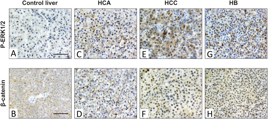
Fig. 6
Deregulation of ERK and Wnt/β-catenin pathways in different types of krasV12-induced liver tumors.
Three types of liver neoplasia including benign HCA, malignant HCC and HB were examined for the expression patterns of P-ERK1/2 and β-catenin via immunohistochemistry. (A,C,E,G) Strong mixed nuclear and cytoplasmic stainings of P-ERK1/2 were detected in three types of krasV12 liver tumors as compared to control liver of non-induced Triple-Tg fish. (B,D,F,H) Immunohistochemistry for β-catenin showed nuclear localization and accumulation of β-catenin only in HCC and HB, whereas normal liver and HCA displayed membranous staining of β-catenin. Scale bars, 50 �M.