Fig. 7
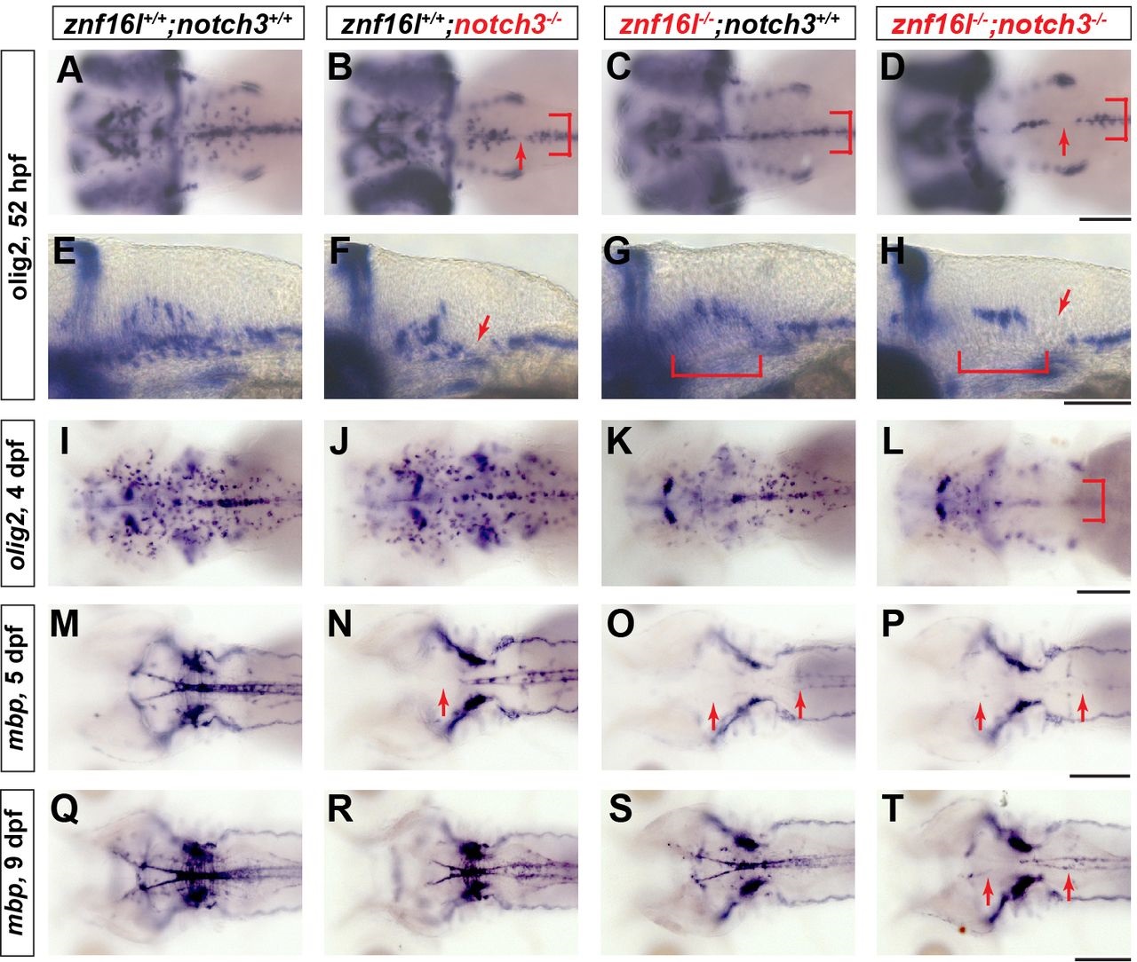
Severe defects in oligodendrocyte development in mutants lacking function of Znf16l and Notch3. In situ hybridization analyses of oligodendrocyte development and myelination in mutants lacking Notch3, Znf16l, or both. (A,E,I,M,Q) Wild-type oligodendrocyte precursors migrating laterally can be detected at 52hpf (A), and strong expression of mbp can be detected at 5dpf (M) continuing to 9dpf (Q). (B,F,J,N,R) notch3 mutants (st51) exhibit reduced lateral migration of OPCs at 52hpf and gaps in olig2 expression (red arrows in B,F and bracket in B). The number and distribution of OPCs recovers by 4dpf (J), although mbp expression is still reduced strongly in hindbrain of notch3 mutants (N). (R) By 9dpf mbp expression recovers, but remains distinguishably reduced compared with wild type. (C,G,K,O,S) znf16l single mutants (st97) show reduction of OPCs at an early stage (bracket in C,G) and a lack of mbp at 5dpf that recovers by 9dpf as shown before (O and S). (D,H,L,P,T) Mutants lacking both Znf16l and Notch3 have a more severe phenotype than either single mutant. Numbers of OPCs are strongly reduced and no OPCs migrate laterally similar to the znf16l mutant at 52hpf (red arrows and bracket in D,H). Numbers of OPCs are still reduced by 4dpf (L). mbp expression in double mutants is strongly reduced at 5dpf (P) and very slight expression is observed by 9dpf (T) compared with either single mutant (R and S). Genotypes of all fish analyzed were determined by PCR assay. Scale bars: 50Ám.

